 Back
BackStudy Guide: The Cardiovascular System II – Blood Vessels (Chapter 18)
Study Guide - Smart Notes
 Tailored notes based on your materials, expanded with key definitions, examples, and context.
The Cardiovascular System II: Blood Vessels
Overview of Blood Vessels
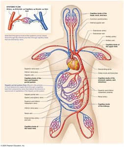
The vasculature is a network of billions of blood vessels that transport blood throughout the body, facilitating the exchange of gases, nutrients, and wastes. Collectively, these vessels measure over 60,000 miles in length.
Arteries: Distribution system, carrying blood away from the heart under high pressure.
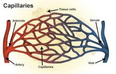
Capillaries: Exchange system, allowing for transfer of substances between blood and tissues.
Veins: Collection system, returning blood to the heart.

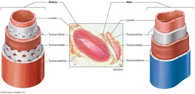
Structure and Function of Arteries and Veins
Blood vessels are composed of three main layers:
Tunica intima: Innermost layer, lined by endothelium.
Tunica media: Middle layer, primarily smooth muscle and elastic fibers.
Tunica externa: Outermost layer, connective tissue.
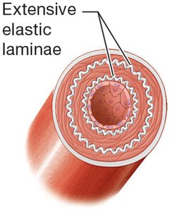
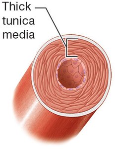
Types of Blood Vessels
Type | Structure | Function |
|---|---|---|
Elastic arteries | Extensive elastic laminae in tunica intima and media | Conduct blood under high pressure |
Muscular arteries | Thick tunica media (smooth muscle) | Control blood flow to organs |
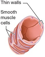
Arterioles | Thin walls, dispersed smooth muscle | Regulate blood pressure, feed capillary beds |
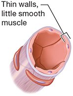
Venules | Thin walls, little smooth muscle | Drain capillary beds |
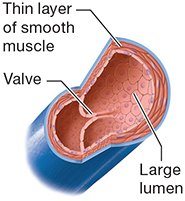
Veins | Thin smooth muscle, large lumen, valves | Return blood to the heart |
Blood Vessel Functions
Conduct blood under high pressure
Control blood flow and regulate blood pressure
Feed and drain capillary beds
Return blood to the heart
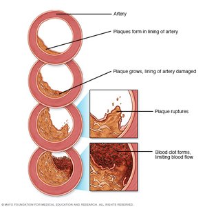
Atherosclerosis
Atherosclerosis is the leading cause of death in the developed world, affecting large and medium-sized arteries. It is characterized by the formation of plaques composed of lipids, cholesterol, calcium salts, and cellular debris within the tunica intima, often at vessel branching points or curves.
Development and Treatment
Injury to endothelium (from high blood pressure, cholesterol, toxins, etc.) leads to inflammation and plaque formation.
Clots may form, causing myocardial infarction or stroke.
Treatment includes lifestyle changes, medications, and possible surgical interventions such as stents or bypass grafts.


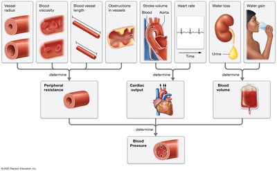
Hemodynamics: Physiology of Blood Flow
Hemodynamics refers to the study of blood flow, which is influenced by pressure and resistance. Blood pressure is the outward force exerted by blood on vessel walls, while resistance is any opposition to blood flow.
High blood pressure can rupture vessel walls; low pressure can cause cell death due to insufficient perfusion.
Key equation: (Pressure = Cardiac Output × Resistance)

Factors Affecting Blood Pressure
Peripheral resistance: Vessel radius, blood viscosity, vessel length
Cardiac output: Heart rate × stroke volume
Blood volume: Influenced by water intake and loss
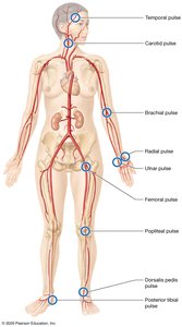
Blood Pressure in Circulation
Systolic Pressure: Pressure during ventricular contraction (~110–120 mm Hg)
Diastolic Pressure: Pressure during ventricular relaxation (~70–80 mm Hg)
Pulse Pressure: Difference between systolic and diastolic (~40 mm Hg)
Mean Arterial Pressure (MAP): Average pressure in systemic arteries during a cardiac cycle.


Pressure in Pulmonary and Systemic Circuits
Circuit | Pressure |
|---|---|
Pulmonary arteries | 15 mm Hg |
Pulmonary veins | 5 mm Hg |
Systemic arteries | 120 mm Hg (systolic), 80 mm Hg (diastolic) |
Arterioles | 80–35 mm Hg |
Capillaries | 35–15 mm Hg |
Venules | 15–5 mm Hg |
Veins | 5–0 mm Hg |
Mechanisms of Venous Return
Valves prevent backflow
Smooth muscle contraction
Skeletal muscle pump
Respiratory pump
Disorders of Blood Pressure
Hypertension
Chronic high blood pressure; "silent killer" due to lack of symptoms
Associated with coronary artery disease, stroke, heart failure, dementia, kidney disease
Treatment: Lifestyle modifications and medications
Hypotension
Low blood pressure; can cause shock, loss of consciousness, organ failure
Causes: Reduced blood volume (hypovolemic shock), decreased cardiac output (cardiogenic shock), excessive vasodilation (anaphylactic or septic shock)
Capillary Structure and Function
Capillaries are the site of exchange between blood and tissues.
Three types: Continuous (least leaky), Fenestrated (moderately leaky), Sinusoidal (leakiest).
Type | Location | Function |
|---|---|---|
Continuous | Skin, nervous tissue, muscle | Permit narrow range of substances |
Fenestrated | Kidneys, endocrine glands, small intestine | Allow large volumes of fluid and larger substances |
Sinusoidal | Liver, bone marrow, spleen | Allow large substances such as cells |
Capillary Exchange Mechanisms
Filtration: Movement of fluid by pressure or gravity
Hydrostatic Pressure (HP): Drives water out of capillary
Osmotic Pressure (OP): Draws fluid into capillary
Net Filtration Pressure (NFP):
Edema
Excessive water in interstitial fluid
Peripheral edema: legs and feet
Ascites: abdomen
Causes: Increased HP, decreased OP, lymphatic obstruction
Disorders: Cerebrovascular Accident (CVA)
Ischemic: Blockage of brain artery (treated with clot-dissolving meds)
Hemorrhagic: Ruptured cerebral artery (treated with surgery)
Risk factors: Hypertension, atherosclerosis, diabetes, smoking, hypercholesterolemia, atrial fibrillation
Symptoms: Sudden paralysis, vision loss, speech difficulty
Summary: The Big Picture of Blood Vessel Anatomy
Blood vessels are essential for distributing blood, maintaining pressure, and facilitating exchange. Disorders such as atherosclerosis, hypertension, and hypotension can significantly impact health, and understanding the structure and function of vessels is crucial for diagnosis and treatment.


Additional info: Some images and tables were inferred for completeness and clarity based on textbook conventions.
