 Back
BackStudy Guide: The Cardiovascular System – The Heart
Study Guide - Smart Notes
 Tailored notes based on your materials, expanded with key definitions, examples, and context.
The Cardiovascular System: The Heart
Heart Anatomy
The heart is a muscular organ responsible for pumping blood throughout the body, delivering oxygen, nutrients, and removing wastes. It is located in the mediastinum, the medial cavity of the thorax, and sits superior to the diaphragm. Most of its mass lies to the left of the midline, and it extends obliquely from the 2nd to the 5th rib.
Size: Approximately the size of a fist (250-350 grams).
Shape: Blunt cone; apex points toward the left side of the body.
Location: Anterior to the vertebral column, posterior to the sternum.
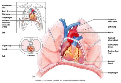
Coverings of the Heart: Pericardium
The heart is enclosed in a double-walled sac called the pericardium. This structure protects, anchors, and reduces friction for the heart during movement.
Fibrous pericardium: Superficial, tough connective tissue.
Serous pericardium: Deep, two-layered membrane (parietal and visceral layers).
Pericardial cavity: Fluid-filled space between layers, containing pericardial fluid.
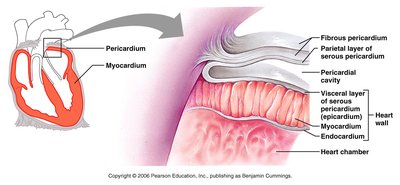
Heart Wall Structure
The heart wall consists of three layers, each with distinct functions:
Epicardium: Visceral layer of serous pericardium, often contains fat.
Myocardium: Cardiac muscle layer, responsible for contraction.
Endocardium: Endothelial layer lining the inner surface.
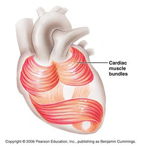
Chambers of the Heart
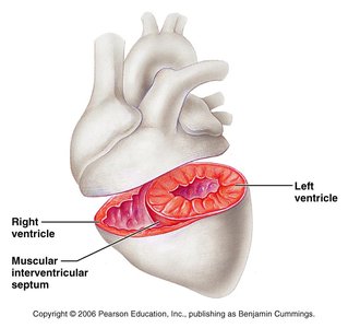
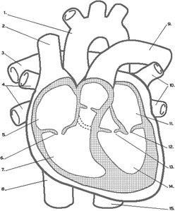
The heart has four chambers: two atria (receiving chambers) and two ventricles (discharging chambers).
Atria: Thin-walled, receive blood returning from circulation. Right atrium receives deoxygenated blood; left atrium receives oxygenated blood.
Ventricles: Thick-walled, pump blood out to pulmonary and systemic circuits.
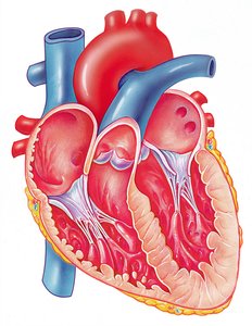
Heart Valves
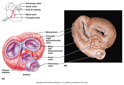
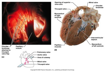
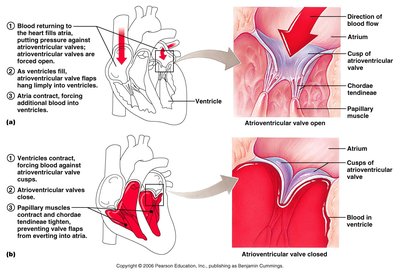
Valves ensure unidirectional blood flow through the heart. There are two types:
Atrioventricular (AV) valves: Between atria and ventricles (tricuspid and bicuspid/mitral).
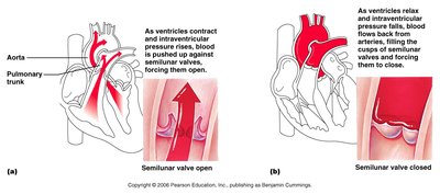
Semilunar (SL) valves: Between ventricles and arteries (aortic and pulmonary).
Flow of Blood Through the Heart
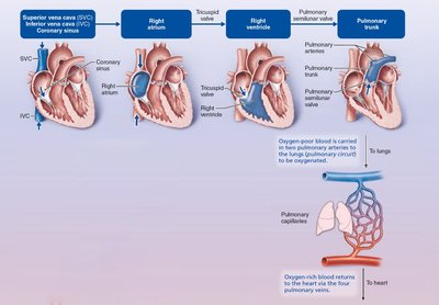
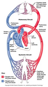
Blood flows through the heart in a specific sequence, ensuring oxygenation and circulation:
Right atrium receives deoxygenated blood from the body.
Blood passes through the tricuspid valve to the right ventricle.
Right ventricle pumps blood through the pulmonary semilunar valve to the pulmonary trunk and arteries.
Blood is oxygenated in the lungs.
Oxygenated blood returns via pulmonary veins to the left atrium.
Blood passes through the bicuspid (mitral) valve to the left ventricle.
Left ventricle pumps blood through the aortic semilunar valve to the aorta.
Blood is distributed to the body via systemic circulation.
Coronary Circulation
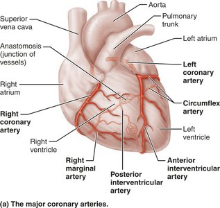
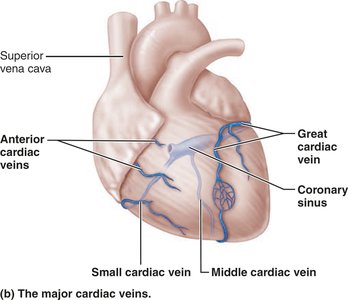
The heart has its own blood supply via the coronary arteries and veins. This circulation is essential for heart muscle function.
Coronary arteries: Arise from the base of the aorta; supply oxygenated blood to the myocardium.
Coronary veins: Collect deoxygenated blood from the myocardium and drain into the coronary sinus.
Anastomoses: Junctions between arteries provide alternate routes for blood delivery.
Microscopic Anatomy of Cardiac Muscle
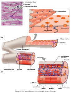
Cardiac muscle is specialized for continuous, rhythmic contraction. It is striated, branched, and interconnected by intercalated discs.
Intercalated discs: Anchor cells and allow ion passage for synchronized contraction.
Functional syncytium: Heart muscle acts as a single unit.
Cardiac Muscle Contraction and Pacemaker Cells
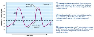
Cardiac muscle contracts in response to action potentials, which are initiated by pacemaker cells. These cells are self-excitable and generate rhythmic impulses.
Pacemaker potential: Slow depolarization due to Na+ influx.
Depolarization: Ca2+ influx triggers action potential.
Repolarization: K+ efflux restores resting potential.
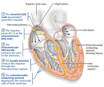
Intrinsic Cardiac Conduction System
The heart's electrical system coordinates contraction. It consists of five main components:
SA (Sinoatrial) Node: Pacemaker, initiates impulse.
AV (Atrioventricular) Node: Delays impulse, allows atrial contraction.
AV Bundle (Bundle of His): Conducts impulse to ventricles.
Bundle Branches: Carry impulse to heart apex.
Purkinje Fibers: Distribute impulse to ventricular walls.
Electrocardiography (ECG/EKG)
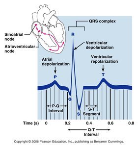
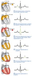
An ECG records the electrical activity of the heart. It consists of three main waves:
P wave: Atrial depolarization.
QRS complex: Ventricular depolarization.
T wave: Ventricular repolarization.
Heart Sounds and Murmurs
Heart sounds are produced by the closing of valves:
Lub: AV valves close (beginning of systole).
Dup: SL valves close (beginning of diastole).
Heart murmurs: Abnormal sounds due to turbulent blood flow, often from valve problems.
Cardiac Cycle
The cardiac cycle includes all events during a heartbeat, marked by pressure and volume changes:
Systole: Contraction phase.
Diastole: Relaxation phase.
Phases: Ventricular filling, isovolumetric contraction, ventricular ejection, isovolumetric relaxation.
Cardiac Output and Regulation
Cardiac output (CO) is the amount of blood pumped by each ventricle per minute. It is calculated as:
CO = HR × SV
HR: Heart rate (beats per minute)
SV: Stroke volume (amount of blood pumped per beat)
Example:
Regulation of Stroke Volume
Stroke volume is determined by:
Preload: Degree of stretch of cardiac muscle before contraction.
Contractility: Force of contraction independent of stretch.
Afterload: Pressure that must be overcome to eject blood.
Formula:
Ejection Fraction:
Regulation of Heart Rate
Heart rate is regulated by:
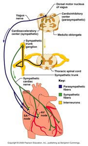
Autonomic nervous system: Sympathetic increases HR, parasympathetic decreases HR.
Chemicals: Hormones (epinephrine, thyroxine) and ions (Ca2+, K+).
Other factors: Age, gender, exercise, body temperature.
Heart rate problems:
Tachycardia: HR > 100 bpm
Bradycardia: HR < 60 bpm
