 Back
BackStudy Guide: The Central Nervous System (CNS) – Structure and Function
Study Guide - Smart Notes
 Tailored notes based on your materials, expanded with key definitions, examples, and context.
The Central Nervous System (CNS)
Overview of the CNS
The Central Nervous System (CNS) is composed of the brain and spinal cord. It is responsible for integrating sensory information and responding accordingly. The evolutionary process known as cephalization led to the development of the anterior portion of the CNS, resulting in a higher concentration of neurons and the complex human brain.
Cephalization: Evolutionary increase in neuron concentration in the head region.
Brain and Spinal Cord: Main components of the CNS.
Brain Development
Embryonic Development of the Brain
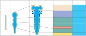
Embryologically, the brain and spinal cord originate from the neural tube. The anterior end of the neural tube expands and forms three primary brain vesicles, which further differentiate into five secondary vesicles, giving rise to the adult brain structures.
Primary Vesicles: Prosencephalon (forebrain), Mesencephalon (midbrain), Rhombencephalon (hindbrain).
Secondary Vesicles: Telencephalon, Diencephalon, Mesencephalon, Metencephalon, Myelencephalon.
Adult Structures: Cerebrum, diencephalon (thalamus, hypothalamus, epithalamus, retina), brain stem (midbrain, pons, medulla oblongata), cerebellum, spinal cord.
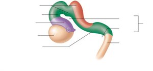
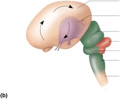
Brain Growth and Folding
The brain grows faster than the skull, resulting in folding to maximize surface area. The forebrain moves toward the brain stem, and the cerebral hemispheres envelop the diencephalon and midbrain.
Brain Regions and Organization
Major Brain Regions
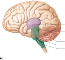
The adult brain is organized into four main regions: cerebral hemispheres, diencephalon, brain stem (midbrain, pons, medulla), and cerebellum.
Cerebral Hemispheres
Diencephalon
Brain Stem: Midbrain, pons, medulla oblongata
Cerebellum
Gray Matter and White Matter Distribution
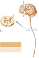
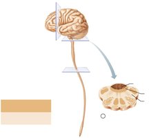
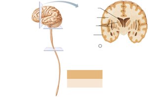
The CNS exhibits a basic pattern: a central cavity surrounded by gray matter, with white matter external to gray matter. This pattern changes as you ascend from the spinal cord to the brain stem and cerebrum.
Gray Matter: Neuron cell bodies, short nonmyelinated neurons.
White Matter: Mostly myelinated axons, some nonmyelinated axons.
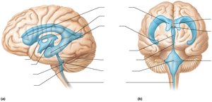
Ventricles of the Brain
Structure and Function
The brain contains fluid-filled chambers called ventricles, which are continuous with each other and the central canal of the spinal cord. They are filled with cerebrospinal fluid (CSF) and lined by ependymal cells.
Lateral Ventricles: Paired, C-shaped chambers in each hemisphere.
Third Ventricle: Located in the diencephalon.
Fourth Ventricle: Located in the hindbrain, continuous with the central canal.
Apertures: Lateral and median apertures connect the fourth ventricle to the subarachnoid space.
Cerebral Hemispheres
Surface Features
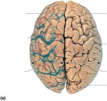
The cerebral hemispheres form the superior part of the brain and account for 83% of its mass. Surface features include gyri (ridges), sulci (shallow grooves), and fissures (deep grooves).
Gyri: Ridges on the brain surface.
Sulci: Shallow grooves separating gyri.
Fissures: Deep grooves, such as the longitudinal fissure (separates hemispheres) and transverse cerebral fissure (separates cerebrum and cerebellum).
Lobes of the Cerebral Hemispheres
Each hemisphere is divided into five lobes: frontal, parietal, temporal, occipital, and insula. The first four are named after the overlying skull bones, while the insula is buried beneath portions of the other lobes.
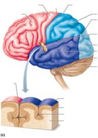
Major Sulci and Regions
Major sulci include the central sulcus (separates frontal and parietal lobes), parieto-occipital sulcus (separates parietal and occipital lobes), and lateral sulcus (outlines temporal lobes). Each hemisphere has three basic regions: cerebral cortex (gray matter), white matter, and basal nuclei.
Cerebral Cortex
Structure and Function
The cerebral cortex is the executive suite of the brain, responsible for conscious mind functions such as awareness, sensory perception, voluntary motor initiation, communication, memory storage, and understanding. It is a thin (2–4 mm) layer of gray matter, making up 40% of the brain's mass.
Neuron Cell Bodies, Dendrites, Glial Cells, Blood Vessels: Components of the cortex.
No Axons: The cortex does not contain axons.
Functional Areas of the Cerebral Cortex
The cortex contains three types of functional areas: motor areas (control voluntary movement), sensory areas (conscious awareness of sensation), and association areas (integrate diverse information). Each hemisphere controls the contralateral side of the body, and lateralization of function can occur in only one hemisphere.
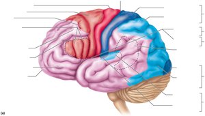
Motor Areas
Motor areas are located in the frontal lobe and include the primary motor cortex (precentral gyrus), premotor cortex, Broca’s area, and frontal eye field.
Primary Motor Cortex: Controls precise, skilled, skeletal muscle movements via pyramidal cells.
Pyramidal (Corticospinal) Tracts: Long axons projecting down the spinal cord.
Somatotopy: Mapping of body muscles to specific cortex areas.
Motor Homunculi: Visual representation of motor innervation.
Premotor Cortex, Broca’s Area, and Frontal Eye Field
Premotor Cortex: Plans movements, controls learned and patterned motor skills, coordinates actions.
Broca’s Area: Motor speech area directs muscles of speech production, usually in the left hemisphere.
Frontal Eye Field: Controls voluntary eye movements.


Sensory Areas
Sensory areas are located in the parietal, insular, temporal, and occipital lobes. They include the primary somatosensory cortex, somatosensory association cortex, visual areas, auditory areas, vestibular cortex, olfactory cortex, gustatory cortex, and visceral sensory area.
Primary Somatosensory Cortex: Receives sensory information from skin and proprioceptors; spatial discrimination.
Somatosensory Association Cortex: Integrates sensory input for object understanding.
Visual Areas: Primary visual cortex and visual association area interpret visual stimuli.
Auditory Areas: Primary auditory cortex and association area interpret sound.
Vestibular Cortex: Awareness of balance.
Olfactory Cortex: Awareness of odors.
Gustatory Cortex: Perception of taste.
Visceral Sensory Area: Perception of visceral sensations.

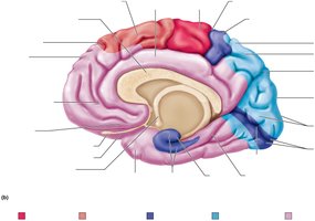
Multimodal Association Areas
Multimodal association areas receive inputs from multiple sensory areas and send outputs to various regions. They allow for meaning, memory storage, and decision-making. The three main parts are the anterior association area (prefrontal cortex), posterior association area, and limbic association area.
Anterior Association Area: Intellect, cognition, recall, personality, judgment, reasoning, planning.
Posterior Association Area: Pattern and face recognition, spatial localization, language understanding (Wernicke’s area).
Limbic Association Area: Emotional impact, memory formation.
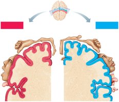
Lateralization of Cortical Functioning
Lateralization refers to the division of labor between hemispheres. The left hemisphere is dominant for language, math, and logic in 90% of humans, while the right hemisphere specializes in visual-spatial skills, intuition, emotion, and artistic abilities. Hemispheres communicate via fiber tracts for functional integration.
Left Hemisphere: Language, math, logic.
Right Hemisphere: Visual-spatial skills, intuition, emotion, artistic and musical skills.
Summary Table: Brain Regions and Functions
Region | Main Function |
|---|---|
Cerebral Hemispheres | Conscious mind, voluntary movement, sensory perception |
Diencephalon | Thalamus, hypothalamus, epithalamus, retina; relay and integration |
Brain Stem | Midbrain, pons, medulla; basic life functions, pathway for signals |
Cerebellum | Coordination of movement, balance |
Key Terms and Definitions
Cephalization: Evolutionary concentration of nervous tissue in the head.
Neural Tube: Embryonic precursor to CNS.
Ventricles: Fluid-filled chambers in the brain.
Gyri/Sulci/Fissures: Surface features of the cerebral cortex.
Gray Matter: Neuron cell bodies and dendrites.
White Matter: Myelinated axons.
Homunculus: Visual representation of body mapping in cortex.
Lateralization: Specialization of hemispheric functions.
Relevant Equations
While the CNS is primarily anatomical, some relevant equations for neurophysiology include:
Nernst Equation: Used to calculate equilibrium potential for ions across a membrane.
Ohm's Law (for neurons): Relationship between voltage, current, and resistance.
