 Back
BackStudy Guide: The Immune System – Innate and Adaptive Body Defenses
Study Guide - Smart Notes
 Tailored notes based on your materials, expanded with key definitions, examples, and context.
CH 21 The Immune System: Overview
General Functions and Structure
The immune system is a functional system that provides resistance to disease-causing microorganisms such as bacteria, fungi, and viruses. It is not an anatomical organ system, but rather a network of molecules and immune cells (especially lymphocytes) that reside in lymphoid tissues and circulate in body fluids. The body employs three lines of defense, acting both independently and cooperatively, to resist invasion and provide immunity.
First Line of Defense: Surface barriers (skin and mucosae)
Second Line of Defense: Innate internal defenses (inflammation, antimicrobial proteins, phagocytes)
Third Line of Defense: Adaptive (specific) defense system (lymphocytes and antibodies)
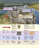
Innate Defenses
Surface Barriers: First Line of Defense
Surface barriers include the skin and mucous membranes, along with their secretions. These barriers are highly effective for most microbes due to their structural and chemical properties.
Keratin: Protein in the epidermis resistant to acids, bases, bacterial enzymes, and toxins
Mucosae: Provide mechanical barriers within the body
Acid Mantle: Acidity of skin, vaginal, and stomach secretions inhibits bacterial growth
Enzymes: Lysozyme in saliva, respiratory mucus, and lacrimal fluid kills microbes; protein-digesting enzymes in stomach destroy pathogens
Mucin: Sticky mucus traps microbes in digestive and respiratory tracts
Defensins: Antimicrobial peptides secreted in response to barrier breach and inflammation
Other Chemicals: Lipids in sebum and dermcidin in sweat are toxic to bacteria
Innate Internal Defenses: Second Line of Defense
If microbes breach the first line, the second line of defense is activated. This includes nonspecific cellular and chemical means such as phagocytes, natural killer cells, antimicrobial proteins, inflammation, and fever.
Pattern Recognition Receptors: Identify pathogens by recognizing specific-shaped molecules (e.g., carbohydrates) found on them
Toll-like Receptors (TLRs): Play a central role in triggering immune responses; humans have 11 TLRs
Phagocytes
Phagocytes are white blood cells that ingest and digest foreign invaders and cellular debris.
Neutrophils: Most abundant phagocytes; phagocytize infectious material in tissues
Macrophages: Most voracious phagocytes; can be free (wandering) or fixed (resident in organs)
Phagocytosis Process
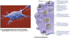
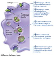
Phagocytosis begins when phagocyte receptors bind to a particle, which is then pulled inside and enclosed within a membrane-lined vesicle (phagosome). The phagosome fuses with a lysosome to form a phagolysosome, where acidification and lysosomal enzymes digest the contents. Some pathogens resist these enzymes, requiring additional mechanisms such as the respiratory burst.
Natural Killer (NK) Cells
NK cells are large granular lymphocytes that police blood and lymph, targeting cells with abnormal surface proteins (e.g., lack of MHC). They kill cancer and virus-infected cells by inducing apoptosis and secrete chemicals that enhance inflammation. 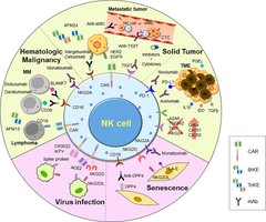
Inflammation
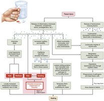
Inflammation is a nonspecific response to tissue injury, caused by trauma, heat, chemicals, or infection.
Benefits: Prevents spread of pathogens, disposes of debris, alerts adaptive immune system, sets stage for repair
Cardinal Signs: Redness, heat, swelling, pain (sometimes impaired function)
Inflammatory Chemicals: Histamine, kinins, prostaglandins, cytokines, complement proteins
Vasodilation: Increases blood flow and capillary permeability, allowing exudate to seep into tissue
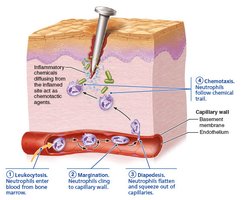
Phagocyte Mobilization
Four steps:
Leukocytosis: Increase in WBCs
Margination: Phagocytes cling to capillary walls
Diapedesis: Neutrophils squeeze between endothelial cells
Chemotaxis: WBCs move toward injured area
Interferons
Interferons are immune-modulating proteins produced by virus-infected cells. They interfere with viral replication in neighboring cells by stimulating production of proteins that block protein synthesis and degrade viral RNA. 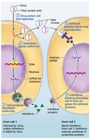
Complement System
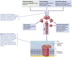
The complement system consists of at least 20 plasma proteins that circulate in an inactive state. When activated, complement amplifies inflammation and lyses certain bacteria and other cell types.
Classical Pathway: Involves antibodies binding to pathogens
Lectin Pathway: Lectins bind to specific sugars on microbes
Alternative Pathway: Spontaneous activation on microbe surfaces
Fever
Fever is a systemic response to invading microbes. Pyrogens released by leukocytes and macrophages act on the hypothalamus to raise body temperature.
Benefits: Enhances immune response, increases metabolic rate, suppresses bacterial growth
Adaptive Defenses
General Features
The adaptive immune system is a specific defensive system that targets and eliminates almost any pathogen. It consists of two overlapping arms:
Humoral Immunity: Antibody-mediated, targets extracellular pathogens
Cellular Immunity: Cell-mediated, targets infected, cancerous, or foreign cells
Antigens
Antigens are substances that trigger the adaptive immune response.
Complete Antigens: Have immunogenicity and reactivity
Haptens: Incomplete antigens that become immunogenic when attached to a protein carrier
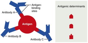
Antigenic Determinants: Parts of antigen that antibodies or lymphocyte receptors bind to
Self-Antigens: MHC Proteins
Self-antigens are proteins on body cell surfaces that are not antigenic to self but are antigenic to others. MHC proteins display peptides (self or foreign) and are essential for T cell recognition. 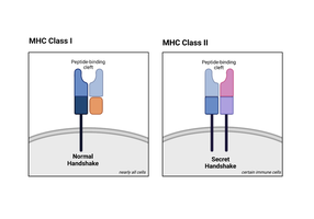
B and T Lymphocytes and Antigen-Presenting Cells
B Lymphocytes: Provide humoral immunity
T Lymphocytes: Provide cellular immunity
Antigen-Presenting Cells (APCs): Present antigens to T cells
Lymphocyte Development, Maturation, and Activation
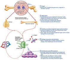
Five general steps:
Origin: Hematopoietic stem cells in red bone marrow
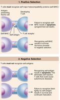
Maturation: Immunocompetence and self-tolerance in primary lymphoid organs
Seeding: Colonization of secondary lymphoid organs
Antigen Encounter and Activation: Clonal selection
Proliferation and Differentiation: Formation of effector and memory cells
Antigen-Presenting Cells (APCs)
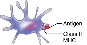
Dendritic Cells: Capture antigens and present them to T cells
Macrophages: Present antigens and become activated phagocytes
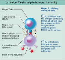
B Lymphocytes: Present antigens to helper T cells
Humoral Immunity
Clonal Selection of B Cells
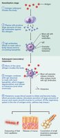
Naive B cells are activated when their surface receptors bind to an antigen, leading to proliferation and differentiation into plasma cells (antibody-secreting) and memory cells. 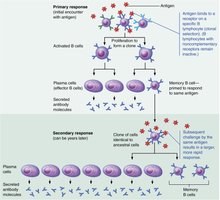
Primary and Secondary Humoral Responses
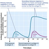
Primary Response: Occurs upon initial exposure; lag period of 3–6 days
Secondary Response: Faster and more effective due to memory cells
Active and Passive Humoral Immunity
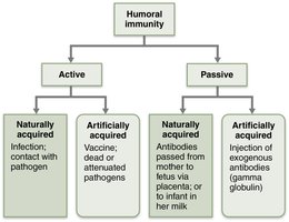
Active Immunity: B cells produce antibodies (naturally via infection or artificially via vaccination)
Passive Immunity: Antibodies are introduced (naturally via placenta/milk or artificially via injection)
Antibodies (Immunoglobulins)
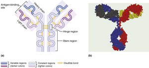
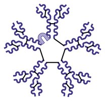
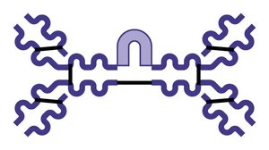
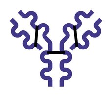
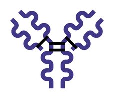
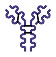
Antibodies are proteins secreted by plasma cells, grouped into five classes: IgG, IgA, IgM, IgE, and IgD.
Structure: Four polypeptide chains (two heavy, two light), variable and constant regions, antigen-binding sites
Classes: Each class has different roles and locations
Antibody Roles
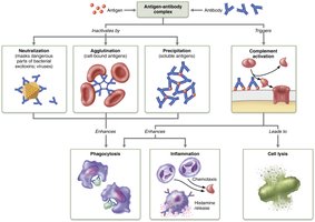
Neutralization: Block sites on pathogens/toxins
Agglutination: Clumping of antigens
Precipitation: Formation of large complexes
Complement Activation: Leads to cell lysis, inflammation, enhanced phagocytosis
Cellular Immunity
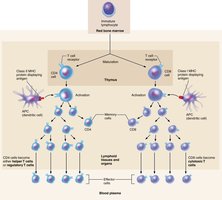
T Lymphocytes
T cells provoke a cellular immune response when presented with antigens.
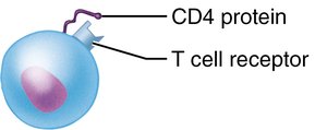
CD4 Cells: Become helper T cells or regulatory T cells
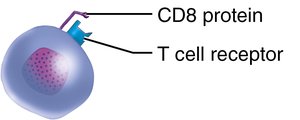
CD8 Cells: Become cytotoxic T cells
MHC Proteins and Antigen Presentation
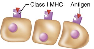
T cells respond only to processed fragments of antigens displayed by MHC proteins.
MHC Class I: On all body cells except RBCs; display endogenous antigens
MHC Class II: On APCs; display exogenous antigens
Activation and Differentiation of T Cells
T cell activation requires antigen binding and co-stimulation. Activated T cells proliferate and differentiate into effector and memory cells. 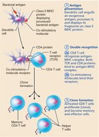
Roles of Specific Effector T Cells
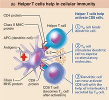
Helper T Cells: Mobilize both humoral and cellular immunity, activate B and T cells, secrete cytokines
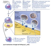
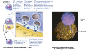
Cytotoxic T Cells: Directly attack and kill infected, cancerous, or foreign cells
Regulatory T Cells: Suppress immune response, prevent autoimmunity
Immune System Disorders
Immunodeficiencies
SCID: Severe combined immunodeficiency; deficit in B and T cells
AIDS: Acquired immune deficiency syndrome; caused by HIV
Autoimmune Diseases
Occur when the immune system fails to distinguish self from foreign antigens, leading to destruction of self tissues. Examples include rheumatoid arthritis, myasthenia gravis, multiple sclerosis, Graves’ disease, type 1 diabetes mellitus, systemic lupus erythematosus, and glomerulonephritis.
Hypersensitivities
Immune responses to perceived threats that cause tissue damage.
Immediate (Acute) Hypersensitivity: Allergies; mediated by IgE
Subacute Hypersensitivity: Mediated by IgM and IgG
Delayed Hypersensitivity: Mediated by T cells
Developmental Aspects of the Immune System
Immune System Development and Aging
Immune system stem cells develop in liver and spleen during early weeks
Bone marrow becomes primary source later
Efficiency wanes with age, increasing susceptibility to infection, cancer, and autoimmune diseases
Thymus atrophies after puberty, reducing naive T and B cell production
Summary Table: Key Differences Between Innate and Adaptive Defenses
Feature | Innate Defenses | Adaptive Defenses |
|---|---|---|
Specificity | Nonspecific | Specific (targets particular antigens) |
Response Time | Immediate | Delayed (requires priming) |
Components | Physical barriers, phagocytes, NK cells, inflammation, complement, fever | B and T lymphocytes, antibodies, APCs |
Memory | No memory | Immunological memory |
Summary Table: Immunoglobulin Classes
Class | Structure | Main Function | Location |
|---|---|---|---|
IgG | Monomer | Main antibody of secondary response; crosses placenta | Blood, lymph |
IgA | Dimer/Monomer | Protects mucosal surfaces | Secretions (saliva, tears, milk) |
IgM | Pentamer | First antibody released; potent agglutinating agent | Blood, lymph |
IgE | Monomer | Triggers histamine release; involved in allergies | Skin, mucosae |
IgD | Monomer | B cell receptor | B cell surface |
Summary Table: Types of Hypersensitivity
Type | Immune Component | Onset | Example |
|---|---|---|---|
Immediate | IgE | Seconds | Allergy, anaphylaxis |
Subacute | IgM, IgG | 1–3 hours | Mismatched blood transfusion |
Delayed | T cells | 1–3 days | Contact dermatitis, TB skin test |
