 Back
BackStudy Guide: The Male Reproductive System (Chapter 27, Marieb Human Anatomy & Physiology)
Study Guide - Smart Notes
 Tailored notes based on your materials, expanded with key definitions, examples, and context.
The Reproductive System: Overview
Functions and Purpose
The reproductive system is responsible for producing offspring and becomes functional at puberty. It carries out four essential tasks:
Formation of gametes: Specialized cells for sexual reproduction—sperm in males and ova (eggs) in females.
Bringing gametes together: Sexual intercourse (copulation) unites male and female gametes.
Fertilization: Fusion of sperm and egg forms a zygote, the first cell of a new organism.
Support of fetal development: Gestation and birth (parturition) are supported by the reproductive system.
Common Features of Male and Female Reproductive Systems
Gonads and Accessory Organs
Male and female reproductive structures share common origins and functions (homologous structures). The primary sex organs, or gonads (testes in males, ovaries in females), produce:
Gametes: Sperm and ova, formed by meiosis.
Sex hormones: Testosterone (males), estrogens and progesterone (females), which regulate organ development, function, and sexual behavior.
Accessory reproductive organs include ducts, glands, and external genitalia.
Hormonal Regulation: The Hypothalamic-Pituitary-Gonadal (HPG) Axis
Hormonal Interactions
The HPG axis regulates gamete and hormone production through a series of hormonal interactions:
GnRH (Gonadotropin-releasing hormone): Released by the hypothalamus, stimulates the anterior pituitary to release gonadotropins.
FSH (Follicle-stimulating hormone) and LH (Luteinizing hormone): Released by the anterior pituitary, act on gonads to stimulate gamete production and hormone secretion.
Sex hormones: Produced by gonads, exert negative feedback on the hypothalamus and anterior pituitary.
Inhibin: Released by gonads, inhibits FSH release at the anterior pituitary.
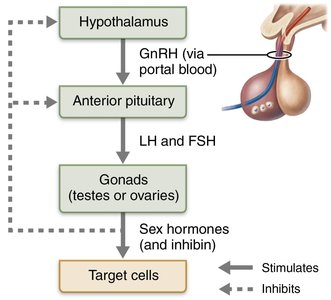
Activation at Puberty
Puberty marks the onset of reproductive function. The hypothalamus becomes less sensitive to inhibition by sex hormones, leading to increased GnRH release, which stimulates FSH and LH production, and subsequently, sex hormone secretion by the gonads.
Anatomy of the Male Reproductive System
Major Structures
The male reproductive system consists of the testes (sperm-producing gonads) and a series of ducts and accessory glands:
Ducts: Epididymis, ductus deferens, ejaculatory duct, urethra
Accessory glands: Seminal glands, prostate, bulbo-urethral glands
External genitalia: Penis, scrotum
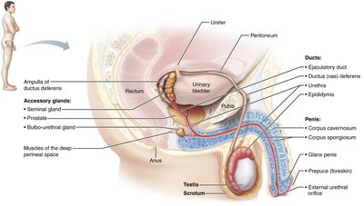
Accessory Glands and Semen Production
Seminal glands: Produce seminal fluid rich in fructose, citric acid, enzymes, and prostaglandins; accounts for ~70% of semen volume.
Prostate: Releases prostatic fluid (milky, slightly acidic, contains citrate and enzymes); activates sperm and accounts for ~30% of semen volume.
Bulbo-urethral glands: Produce mucus that lubricates the glans penis and neutralizes acidic urine in the urethra.
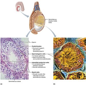
Physiology of the Male Reproductive System
Spermatogenesis: Formation of Sperm
Spermatogenesis is the process of forming sperm in the seminiferous tubules, beginning at puberty and continuing throughout life. Healthy adult males produce approximately 90 million sperm daily.
Histology of the Seminiferous Tubules
Each seminiferous tubule contains four key cell types:
Sustentocytes (nurse cells): Support and nourish developing sperm cells.
Spermatogenic cells: Develop into sperm.
Myoid cells: Smooth muscle-like cells that help move sperm and fluid through the tubules.
Interstitial endocrine cells (Leydig cells): Produce testosterone and small amounts of estrogen.
Phases of Spermatogenesis
Mitosis of spermatogonia: Maintains stem cell line; produces Type A (stem cell) and Type B (primary spermatocyte) daughter cells.
Meiosis: Primary spermatocytes undergo meiosis I and II to form four spermatids.
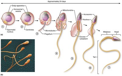
Spermiogenesis: Spermatids transform into mature sperm by elongating, losing excess cytoplasm, and forming a tail.
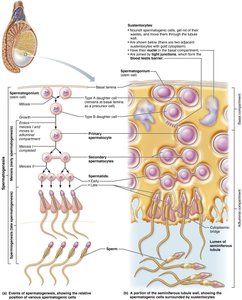
Spermiogenesis: Transformation of Spermatids
Head: Contains nucleus and acrosome (enzymes for egg penetration).
Midpiece: Contains mitochondria for ATP production.
Tail (flagellum): Provides motility.
Role of Sustentocytes (Nurse Cells)
Envelope developing spermatogenic cells and form the blood-testis barrier via tight junctions.
Divide tubule into basal (diploid cells, recognized as self) and adluminal (haploid cells, protected from immune system) compartments.
Prevent immune response against new antigens on spermatogenic cells.
Hormonal Regulation of Male Reproductive Function
HPG Axis in Testicular Function
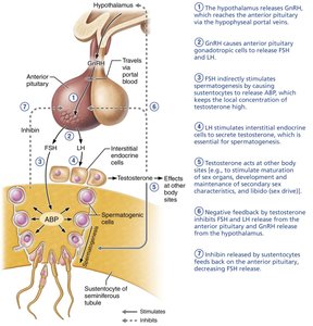
GnRH: Stimulates anterior pituitary to release FSH and LH.
FSH: Stimulates sustentocytes to release androgen binding protein (ABP), maintaining high local testosterone for spermatogenesis.
LH: Stimulates Leydig cells to secrete testosterone.
Testosterone and inhibin: Provide negative feedback to hypothalamus and anterior pituitary; testosterone inhibits GnRH, FSH, and LH, while inhibin inhibits only FSH.
Testosterone: Stimulates maturation of sex organs, secondary sex characteristics, and libido.
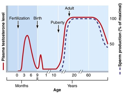
Testosterone Timeline and Effects
Elevated levels before birth for development of male structures; low levels during childhood; rise at puberty to establish adult hormone pattern.
Balance of HPG hormones takes ~3 years to achieve; stable testosterone and sperm production until late in life.
Without GnRH and gonadotropins, testes atrophy and sperm/testosterone production ceases.
Mechanism and Effects of Testosterone
Testosterone: Activates genes to regulate protein synthesis; may be converted to dihydrotestosterone (DHT) or estradiol in target tissues.
Promotes spermatogenesis, growth/maturation of accessory organs, and development of secondary sex characteristics (hair growth, deepening voice, increased bone/muscle mass, increased basal metabolic rate, libido).
Deficiency leads to atrophy of accessory organs, reduced semen volume, and impaired erection/ejaculation; treated with testosterone replacement therapy.
Summary Table: Key Male Reproductive Structures and Functions
Structure | Main Function |
|---|---|
Testes | Sperm production, testosterone secretion |
Epididymis | Sperm maturation and storage |
Ductus deferens | Transport of sperm |
Seminal glands | Produce seminal fluid (70% of semen) |
Prostate | Produce prostatic fluid (activates sperm) |
Bulbo-urethral glands | Produce mucus (lubrication, neutralization) |
Penis | Copulation, delivery of sperm |
Summary Table: Hormonal Regulation of Spermatogenesis
Hormone | Source | Function |
|---|---|---|
GnRH | Hypothalamus | Stimulates FSH and LH release |
FSH | Anterior pituitary | Stimulates sustentocytes, ABP production |
LH | Anterior pituitary | Stimulates Leydig cells, testosterone production |
Testosterone | Testes (Leydig cells) | Promotes spermatogenesis, secondary sex characteristics |
Inhibin | Testes (sustentocytes) | Inhibits FSH release |
Key Equations and Concepts
Meiosis in Spermatogenesis
Meiosis reduces chromosome number by half, producing haploid gametes:
Negative Feedback Regulation
Testosterone and inhibin regulate hormone levels via negative feedback:
Example: Hormonal Regulation in Puberty
As puberty approaches, the hypothalamus releases GnRH in pulses, stimulating the anterior pituitary to release FSH and LH. These hormones act on the testes to increase testosterone production, which in turn promotes spermatogenesis and the development of secondary sex characteristics.
Additional info:
Homologous structures in male and female reproductive systems (e.g., testes and ovaries, penis and clitoris) share developmental origins and functions.
The blood-testis barrier is essential for protecting developing sperm from immune attack.
Testosterone's somatic effects include increased bone and muscle mass, and its behavioral effects include increased libido.
