 Back
Study Guide: The Nervous System (Organization, Cells, and Functional Anatomy)
Study Guide - Smart Notes
 Tailored notes based on your materials, expanded with key definitions, examples, and context.
Organization and Cells of the Nervous System
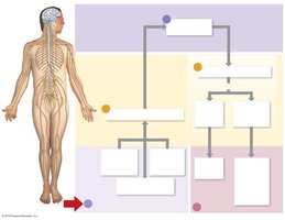
Organization of the Nervous System
The nervous system is divided into two main parts: the central nervous system (CNS) and the peripheral nervous system (PNS). Each plays a distinct role in processing and transmitting information throughout the body.
Central Nervous System (CNS): Consists of the brain and spinal cord. Responsible for integration, processing, and coordination of sensory data and motor commands.
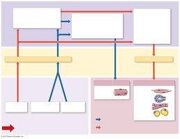
Peripheral Nervous System (PNS): Includes cranial nerves, spinal nerves, and all neural tissue outside the CNS. Divided into sensory (afferent) and motor (efferent) divisions.
Sensory Division: Receptors collect information and send it to the CNS. Types include somatic sensory receptors (touch, pain, temperature), visceral sensory receptors (internal organs), and special sensory receptors (smell, taste, vision, hearing, balance).
Motor Division: Sends signals to effectors (target organs). Subdivided into the somatic nervous system (SNS, voluntary control of skeletal muscle) and autonomic nervous system (ANS, involuntary control of smooth/cardiac muscle, glands, adipose tissue). The ANS is further divided into sympathetic (fight or flight) and parasympathetic (rest and digest) branches.
Neurons: Structure and Function
Neurons are specialized cells that receive and transmit electrical impulses. Their structure is adapted for rapid communication.
Cell Body: Contains the nucleus and organelles.
Dendrites: Branched extensions that receive signals.
Axon: Long, singular process that transmits impulses away from the cell body.
Axon Hillock: Transition region from cell body to axon; site of action potential initiation.
Axon Terminal: Contains vesicles of neurotransmitter chemicals for communication with other cells.
Structural Types of Neurons
Neurons are classified by their structure:
Multipolar Neurons: Many dendrites, one axon; most common in CNS and motor neurons.
Bipolar Neurons: One dendrite, one axon; found in special sensory organs.
Unipolar Neurons: Single process that splits into two branches; most sensory neurons.
Neuroglia (Glial Cells)
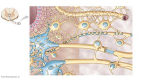
Neuroglia are support cells in the CNS and PNS, accounting for about half the volume of the nervous system.
Astrocytes: Form the blood-brain barrier and provide structural support.
Microglia: Phagocytic defense cells.
Ependymal Cells: Produce and circulate cerebrospinal fluid (CSF).
Oligodendrocytes: Create the myelin sheath in the CNS.
Myelin and Neuroglia in the PNS
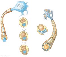
Myelin is a lipid-rich substance that wraps around axons, speeding up conduction.
Schwann Cells: Provide myelination in the PNS; their outer surface is called the neurilemma.
Satellite Cells: Surround cell bodies in the PNS, forming ganglia.
Myelinated Axons: Form white matter; unmyelinated axons and cell bodies/dendrites make up gray matter.
Functional Classes of Neurons
Neurons are also classified by function:
Sensory Neurons: Unipolar; transmit information from receptors to CNS via afferent fibers.
Interneurons: Multipolar; located in CNS, responsible for higher functions (learning, memory, planning).
Motor Neurons: Multipolar; carry information from CNS to effectors via efferent fibers. Somatic motor neurons innervate skeletal muscle; visceral motor neurons innervate cardiac/smooth muscle, glands, adipose tissue.
Neuron Physiology: An Electrochemical Event
Membrane Potential
Neurons maintain a polarized membrane at rest, with a positive charge outside and negative charge inside due to proteins and ion distribution.
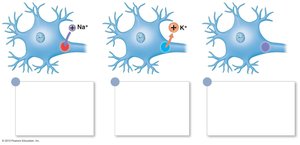
Ion Channels: Allow diffusion of ions; can be leak (always open), ligand-gated (open when bound to neurotransmitter), or voltage-gated (open in response to electrical changes).
Resting Potential: The normal, polarized state of the neuron.
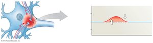
Graded Potential: Local change in membrane potential caused by a stimulus.
Action Potential: If graded potential is large enough, an action potential develops and propagates down the axon.
Propagation of Action Potentials
Action potentials travel along axons by two methods:
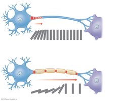
Continuous Propagation: Occurs in unmyelinated axons; action potential moves stepwise along the entire membrane.
Saltatory Propagation: Occurs in myelinated axons; action potential jumps from node to node (nodes of Ranvier), increasing speed.
Synapse and Neurotransmitters
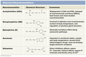
The synapse is the site of communication between neurons, using neurotransmitters.
Neurotransmitter Release: Action potential opens calcium channels at the axon terminal, triggering exocytosis of neurotransmitter vesicles.
Binding: Neurotransmitters bind to receptors on the postsynaptic cell, causing graded potentials.
Removal: Neurotransmitters are broken down enzymatically or reabsorbed by the axon terminal.
Functional Anatomy of the Brain
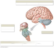
Major Regions of the Brain
The brain is organized into several major regions, each with specialized functions.
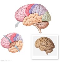
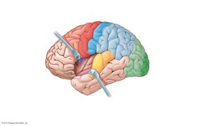
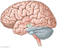
Cerebrum: Divided into two hemispheres; responsible for conscious thought, memory, sensory processing, and voluntary motor control. Surface features include gyri (ridges), sulci (grooves), and fissures (deep grooves).
Cerebellum: Coordinates motor commands and maintains balance.
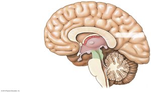
Diencephalon: Includes thalamus (sensory relay) and hypothalamus (emotions, autonomic functions, hormone control).
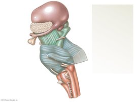
Brain Stem: Includes midbrain (visual/auditory reflexes), pons (relay centers, motor control), and medulla oblongata (vital autonomic functions).
Brain Organization: Gray and White Matter
Nuclei: Clusters of cell bodies (gray matter) where processing occurs.
Tracts: Bundles of axons (white matter) for signal transmission.
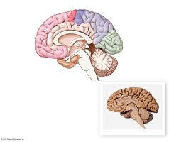
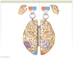
Corpus Callosum: Major tract connecting cerebral hemispheres.
Basal Nuclei: Islands of gray matter within deeper white matter.
Four Lobes of the Cerebrum
Each lobe of the cerebrum has specialized functions:
Frontal Lobe: Prefrontal cortex (abstract functions), somatic motor association area, primary motor cortex, gustatory cortex, Broca's area (speech).
Parietal Lobe: Primary sensory cortex, somatic sensory association area, spatial analysis.
Temporal Lobe: Olfactory cortex (smell), auditory cortex (hearing), general interpretive area.
Occipital Lobe: Primary visual cortex, visual association area.
Diencephalon
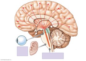
Thalamus: Final relay point for all senses except olfaction; passes information to relevant cortices and subconscious centers.
Hypothalamus: Controls emotions, drives (hunger, thirst), body temperature, coordinates nervous/endocrine activities, produces hormones (ADH, oxytocin).
Pineal Gland: Produces melatonin, regulating circadian rhythms.
Brain Stem
Midbrain: Processes visual and auditory data, generates reflexes, maintains consciousness.
Pons: Sensory relay to cerebellum and thalamus, motor centers for breathing.
Medulla Oblongata: Sensory relay, autonomic centers for cardiovascular, respiratory, and digestive functions.
Cerebrospinal Fluid and Ventricles
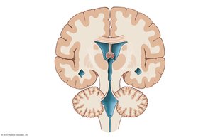
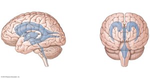
The brain contains internal chambers called ventricles, filled with cerebrospinal fluid (CSF) produced by choroid plexuses.
Lateral Ventricles: Inside cerebral hemispheres.
Third Ventricle: In diencephalon.
Cerebral Aqueduct: Through midbrain.
Fourth Ventricle: In pons and medulla oblongata.
CSF: Bathes the brain, acts as a shock absorber.
Meninges
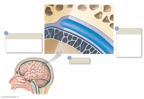
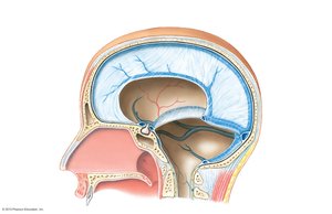
The meninges are protective layers surrounding the brain and spinal cord.
Dura Mater: Outermost, tough and fibrous; contains dural sinuses with venous blood.
Arachnoid Mater: Middle layer; subarachnoid space holds CSF.
Pia Mater: Innermost; follows all folds and blood vessels.
Cerebrospinal Fluid Production and Circulation
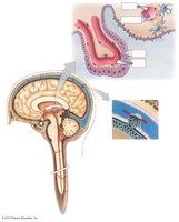
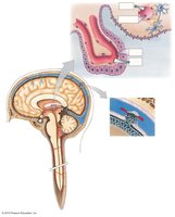
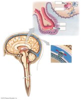
Choroid Plexuses: Produce CSF from capillaries.
CSF Circulation: Travels out of ventricles, down central canal or into subarachnoid space.
Removal: CSF is removed from subarachnoid space through arachnoid granulations into dural sinuses.
Additional info:
Neurotransmitters play a critical role in synaptic transmission, with different types having excitatory or inhibitory effects.
Action potentials are generated by rapid changes in membrane potential, primarily due to sodium and potassium ion movement.
Myelination increases the speed of action potential propagation, which is essential for rapid communication in high-priority sensory and motor pathways.
