 Back
Study Guide: The Reproductive System (Chapter 27) – Human Anatomy & Physiology
Study Guide - Smart Notes
 Tailored notes based on your materials, expanded with key definitions, examples, and context.
The Reproductive System: Overview
General Functions and Organization
The reproductive system is responsible for producing offspring and ensuring the continuation of genetic material. It consists of primary sex organs (gonads) that produce gametes and sex hormones, and accessory organs that support reproduction.
Gonads: Testes in males, ovaries in females; produce gametes (sperm or ova) and sex hormones.
Accessory Organs: Structures that transport, nourish, or protect gametes and support gestation.
Four Main Tasks:
Form gametes for sexual reproduction.
Bring gametes together via copulation.
Combine genetic information through fertilization (formation of zygote).
Support fetal development and birth.
Common Features of Male and Female Reproductive Systems
Gonads and Hormonal Regulation
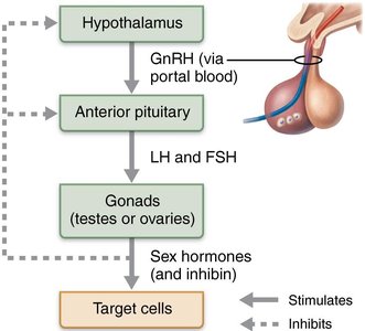
Both male and female reproductive systems share homologous structures and are regulated by the hypothalamic-pituitary-gonadal (HPG) axis.
HPG Axis: Hormonal cascade involving GnRH (from hypothalamus), FSH and LH (from anterior pituitary), and steroid hormones (from gonads).
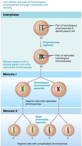
Meiosis: Specialized cell division that reduces chromosome number by half and introduces genetic variability.
Meiosis: Basis of Gamete Production
Overview and Unique Events
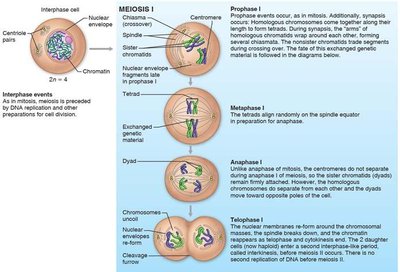
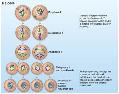
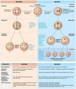
Meiosis consists of two consecutive nuclear divisions (Meiosis I and II) without DNA replication in between, resulting in four genetically unique haploid cells.
Synapsis: Homologous chromosomes pair up.
Crossing Over: Exchange of genetic material between homologous chromosomes.
Reduction Division: Chromosome number is halved.
Comparison of Mitosis and Meiosis
Mitosis produces two identical diploid cells, while meiosis produces four genetically distinct haploid cells.
Mitosis: Occurs in somatic cells; for growth and repair.
Meiosis: Occurs in gametes; for sexual reproduction.
Anatomy of the Male Reproductive System
Testes and Duct System
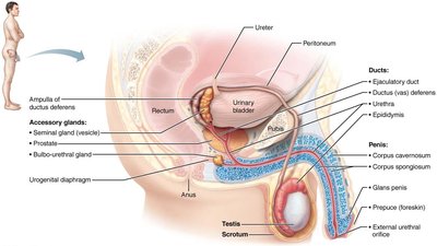
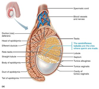
The testes are the primary male gonads, producing sperm and testosterone. Sperm is transported through a series of ducts and supported by accessory glands.
Testes: Located in the scrotum; contain seminiferous tubules for sperm production.
Ducts: Epididymis, ductus deferens, ejaculatory duct, urethra.
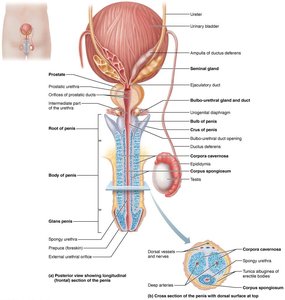
Accessory Glands: Seminal glands, prostate, bulbo-urethral glands.
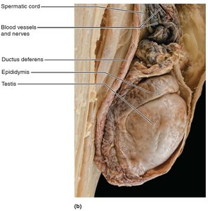
Scrotum and Testes Structure
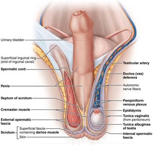
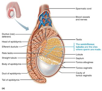
The scrotum maintains a temperature lower than body temperature, essential for viable sperm production. Each testis is divided into lobules containing seminiferous tubules.
Tunica Albuginea: Dense connective tissue covering the testis.
Lobules: Each contains seminiferous tubules for spermatogenesis.
Male Duct System
The duct system transports sperm from the testes to the exterior.
Epididymis: Site of sperm maturation and storage.
Ductus (Vas) Deferens: Propels sperm during ejaculation.
Urethra: Conducts semen and urine to the exterior.
Penis and Male Perineum
Structure and Function
The penis is composed of erectile tissue and is involved in copulation. The male perineum supports the scrotum and penis.
Corpus Spongiosum and Corpora Cavernosa: Erectile tissues responsible for erection.
Perineum: Region bounded by pubic symphysis, ischial tuberosities, and coccyx.
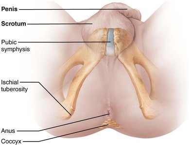
Male Accessory Glands and Semen Composition
Accessory Glands
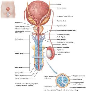
Accessory glands produce seminal fluid, which nourishes and protects sperm.
Seminal Glands: Provide fructose and prostaglandins.
Prostate: Activates sperm and provides enzymes.
Bulbo-Urethral Glands: Secrete mucus for lubrication and neutralization.
Semen Composition and Function
Semen: Alkaline fluid containing sperm, nutrients, enzymes, and protective chemicals.
Key Functions: Energy supply (fructose), sperm activation (alkaline pH, relaxin), motility, protection (antibiotics, immune suppression), coagulation and liquefaction.
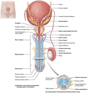
Male Sexual Response
Erection and Ejaculation
Erection is controlled by parasympathetic reflexes, while ejaculation is a sympathetic reflex.
Erection: Vasodilation of penile arterioles via nitric oxide (NO).
Ejaculation: Propulsion of semen via emission and expulsion phases.
Orgasm: Includes ejaculation and CNS-mediated pleasurable sensations.
Refractory Period: Recovery interval post-ejaculation.
Spermatogenesis
Process and Cell Types
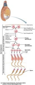
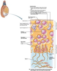
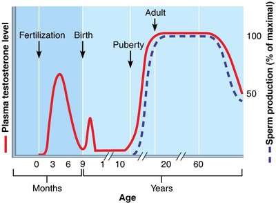
Spermatogenesis is the production of male gametes in the seminiferous tubules, beginning at puberty and continuing throughout life.
Spermatogonia: Stem cells; divide by mitosis.
Primary Spermatocytes: Undergo meiosis I.
Secondary Spermatocytes: Undergo meiosis II.
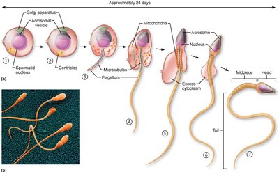
Spermatids: Haploid, nonfunctional.
Spermatozoa: Mature, motile gametes.
Spermiogenesis: Transformation of spermatids into functional sperm.
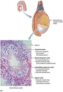
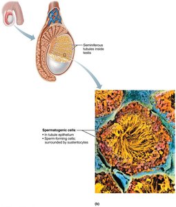
Sustentocytes and Interstitial Endocrine Cells
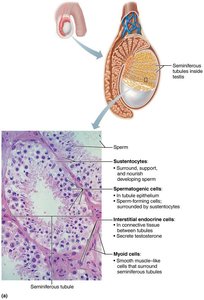
Sustentocytes (Sertoli cells) support and nourish developing sperm, form the blood-testis barrier, and secrete fluid for sperm transport. Interstitial endocrine cells (Leydig cells) produce testosterone.
Sustentocytes: Stimulated by FSH; produce ABP and inhibin.
Interstitial Endocrine Cells: Stimulated by LH; produce testosterone.
Regulation of Male Reproductive System
Hormonal Control (HPG Axis)
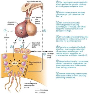
The HPG axis regulates spermatogenesis and testosterone production via feedback loops.
GnRH: Stimulates FSH and LH release.
FSH: Stimulates sustentocytes to produce ABP.
LH: Stimulates interstitial cells to produce testosterone.
Testosterone: Inhibits GnRH and LH release.
Inhibin: Inhibits FSH release.
Anatomy of the Female Reproductive System
Organs and Structure
The female reproductive system produces gametes and sex hormones, and supports gestation.
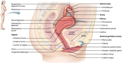
Ovaries: Flank the uterus; contain follicles at various stages.
Uterine Tubes: Transport oocytes; fimbriae and peristalsis aid movement.
Uterus: Fundus, body, cervix; wall consists of perimetrium, myometrium, endometrium.
Vagina: Copulatory organ; passage for menstrual flow and childbirth.
Oogenesis
Process and Timeline
Oogenesis is the production of eggs, beginning before birth and continuing through reproductive life.
Oogonia: Diploid stem cells; become primary oocytes before birth.
Primary Oocytes: Arrested in prophase I until puberty.
Secondary Oocyte: Produced monthly; completes meiosis II only if fertilized.
Polar Bodies: Nonfunctional byproducts of meiosis.
Comparison: Spermatogenesis vs Oogenesis
Oogenesis yields one functional ovum per primary oocyte, while spermatogenesis yields four functional sperm.
Process | Number of Gametes | Timing |
|---|---|---|
Spermatogenesis | 4 sperm per primary spermatocyte | Continuous from puberty |
Oogenesis | 1 ovum per primary oocyte | Begins before birth, resumes at puberty |
The Ovarian Cycle
Phases and Follicle Development
The ovarian cycle consists of follicular and luteal phases, with ovulation occurring mid-cycle.
Follicular Phase (Days 1–14): Follicle growth and maturation; dominant follicle selected.
Ovulation (Day 14): Secondary oocyte released.
Luteal Phase (Days 15–28): Corpus luteum produces hormones; degenerates if no pregnancy.
Regulation of Female Reproductive System
Hormonal Control of the Ovarian Cycle
The ovarian cycle is regulated by a hormonal cascade involving GnRH, FSH, LH, estrogens, and progesterone, with feedback mechanisms controlling cycle progression.
GnRH: Stimulates FSH and LH release.
FSH: Stimulates follicle growth and estrogen production.
LH: Triggers ovulation and corpus luteum formation.
Estrogen: Negative feedback at low levels; positive feedback at high levels (LH surge).
Progesterone: Maintains luteal phase; strong negative feedback.
Summary Table: Key Differences Between Male and Female Gametogenesis
Feature | Spermatogenesis | Oogenesis |
|---|---|---|
Location | Seminiferous tubules (testes) | Ovarian follicles (ovaries) |
Timing | Puberty to death | Before birth to menopause |
Yield | 4 sperm per primary spermatocyte | 1 ovum per primary oocyte |
Cell Division | Equal | Unequal (cytoplasm concentrated in ovum) |
