 Back
BackStudy Guide: The Skeletal System (Essentials of Human Anatomy & Physiology, Ch. 5)
Study Guide - Smart Notes
 Tailored notes based on your materials, expanded with key definitions, examples, and context.
The Skeletal System
Overview and Parts of the Skeletal System
The skeletal system is a complex framework of bones, joints, cartilages, and ligaments that provides structure and support to the human body. It is divided into two main subdivisions: the axial skeleton and the appendicular skeleton.
Bones (skeleton): The rigid organs forming the main structure.
Joints: Articulations where two or more bones meet.
Cartilages: Flexible connective tissue found in joints and other areas.
Ligaments: Tough bands connecting bones at joints.
Axial skeleton: Includes the skull, vertebral column, and thoracic cage.
Appendicular skeleton: Includes limbs and girdles (shoulder and pelvic).
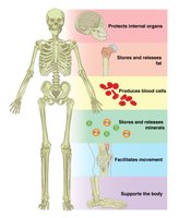
Functions of Bones
Bones serve several essential functions in the body, including support, protection, movement, storage, and blood cell formation.
Support: Provides structural framework for the body.
Protection: Shields vital organs (e.g., skull protects brain, rib cage protects thoracic organs).
Movement: Facilitates movement through attached muscles.
Storage: Stores minerals (calcium, phosphorus) and fats (in marrow).
Blood cell formation (hematopoiesis): Occurs in bone marrow.
Classification of Bones
Types of Osseous Tissue
Bones are composed of two main types of tissue:
Compact bone: Dense, smooth, and forms the outer layer.
Spongy bone: Contains trabeculae (needlelike structures) and open spaces.
Bone Classification by Shape
Bones are classified into four groups based on their shape:
Long bones: Longer than wide, mostly compact bone; found in limbs (e.g., femur, humerus).
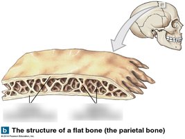
Flat bones: Thin, flattened, usually curved; two layers of compact bone sandwiching spongy bone (e.g., skull, ribs, sternum).
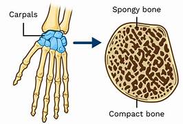
Short bones: Cube-shaped, mostly spongy bone (e.g., carpals, tarsals).
Irregular bones: Complex shapes, do not fit other categories (e.g., vertebrae, hip bones).
Structure of Bone
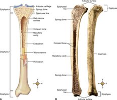
Long Bone Anatomy
Long bones have a distinct structure, including the diaphysis, epiphysis, and various membranes.
Diaphysis: Shaft, composed of compact bone.
Epiphysis: Ends, mostly spongy bone.
Periosteum: Outer covering of diaphysis, fibrous connective tissue.
Articular cartilage: Covers epiphyses, reduces friction.
Epiphyseal plate/line: Growth plate in young bones; line in adults.
Endosteum: Lines inner surface of shaft.
Medullary cavity: Contains yellow marrow (fat) in adults.
Bone Markings
Bone markings are features on bones that serve as sites for muscle, tendon, and ligament attachment, or as passages for nerves and blood vessels.
Projections/processes: Grow out from bone surface (begin with "T").
Depressions/cavities: Indentations (begin with "F", except facet).
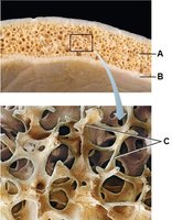
Microscopic Anatomy of Bone
Spongy bone: Contains trabeculae and open spaces filled with marrow, blood vessels, and nerves.
Compact bone: Contains osteocytes (mature bone cells) in lacunae (cavities), arranged in lamellae (concentric circles) around a central (Haversian) canal.
Osteon (Haversian system): Structural unit of compact bone.
Canaliculi: Tiny canals connecting osteocytes to nutrient supply.
Bone Composition
Bone is lightweight yet strong, due to its organic (collagen fibers) and inorganic (calcium salts) components.
Collagen fibers: Provide flexibility and tensile strength.
Calcium salts: Provide hardness and resistance to compression.
Bone Formation, Growth, and Remodeling
Ossification and Growth
Ossification is the process of bone formation, occurring on hyaline cartilage models or fibrous membranes. Long bone growth involves two major phases:
Osteoblasts cover cartilage model with bone matrix.
Cartilage is digested away, opening up a medullary cavity.
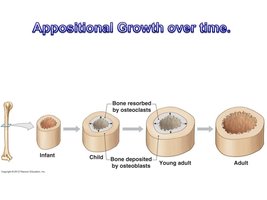
Appositional Growth
Bones grow in width through appositional growth, where osteoblasts add bone matrix to the outside and osteoclasts remove bone from the inside. Growth is regulated by hormones.
Growth hormone: Stimulates bone growth.
Sex hormones: Influence bone growth during puberty.
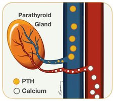
Calcium Ion Regulation
Calcium levels in the blood are regulated by the parathyroid hormone (PTH).
PTH: Released when blood calcium is low; activates osteoclasts to break down bone and release calcium.
Axial Skeleton
Components of the Axial Skeleton
The axial skeleton forms the longitudinal axis of the body and includes:
Skull: Protects the brain.
Vertebral column: Provides support and protection for the spinal cord.
Bony thorax: Protects thoracic organs.
Skull
Cranium: Encloses the brain (8 bones).
Facial bones: Hold eyes and allow facial expressions (14 bones).
Sutures: Immovable joints between skull bones.
Mandible: Only freely movable joint in the skull.
Paranasal sinuses: Hollow portions that lighten the skull and amplify sounds.
Hyoid bone: Only bone not articulating with another; aids in tongue movement and speech.
Vertebral Column
26 vertebral bones: Separated by intervertebral discs.
Cervical (7), thoracic (12), lumbar (5), sacrum (5 fused), coccyx (3–5 fused).
Primary curvatures: Thoracic and sacral (C-shaped in newborns).
Secondary curvatures: Cervical and lumbar (S-shaped in adults).
Common features: Body, vertebral arch, pedicle, lamina, vertebral foramen, transverse and spinous processes, articular processes.
Thoracic Cage
Sternum: Central bone of the chest.
Ribs: 12 pairs (true, false, floating).
Thoracic vertebrae: Posterior attachment for ribs.
Appendicular Skeleton
Components of the Appendicular Skeleton
The appendicular skeleton consists of 126 bones, including limbs and girdles.
Pectoral girdle: Clavicle and scapula; attaches upper limb to axial skeleton.
Pelvic girdle: Coxal bones, sacrum, coccyx; supports lower limb and protects organs.
Bones of the Upper Limbs
Humerus: Arm bone; articulates with scapula and forearm bones.
Ulna: Medial forearm bone; articulates with humerus.
Radius: Lateral forearm bone; articulates with humerus.
Carpals: 8 wrist bones.
Metacarpals: 5 palm bones.
Phalanges: 14 finger bones.
Bones of the Pelvic Girdle
Ilium, ischium, pubis: Fused bones forming coxal bone.
Pelvis: Coxal bones, sacrum, coccyx; supports body weight and protects organs.
Female pelvis: Larger inlet, shallower, lighter, more rounded pubic arch.
Bones of the Lower Limbs
Femur: Thigh bone; strongest bone.
Tibia: Shinbone; forms knee joint.
Fibula: Lateral to tibia; forms outer ankle.
Tarsals: 7 ankle bones (calcaneus, talus).
Metatarsals: 5 bones forming sole.
Phalanges: 14 toe bones.
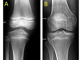
Joints
Overview of Joints
Joints, or articulations, are sites where two or more bones meet. They serve to hold bones together securely and allow for mobility.
Types of joints: Classified by structure and function (not detailed in this excerpt).
Summary Table: Bone Classification by Shape
Type | Description | Examples |
|---|---|---|
Long | Longer than wide, mostly compact bone | Femur, humerus |
Flat | Thin, flattened, usually curved; two layers of compact bone sandwiching spongy bone | Skull, ribs, sternum |
Short | Cube-shaped, mostly spongy bone | Carpals, tarsals |
Irregular | Complex shape, does not fit other categories | Vertebrae, hip bones |
Key Equations and Terms
Ossification: Process of bone formation.
Hematopoiesis: Formation of blood cells in bone marrow.
Appositional growth: Increase in bone width.
Parathyroid hormone (PTH): Regulates blood calcium levels.
Relevant Formula (Calcium Regulation)
Parathyroid hormone increases blood calcium by stimulating osteoclasts:
Additional info: This formula summarizes the hormonal regulation of calcium homeostasis in bone.
