 Back
BackStudy Notes: Blood (Chapter 17) – Human Anatomy & Physiology
Study Guide - Smart Notes
 Tailored notes based on your materials, expanded with key definitions, examples, and context.
Blood: The Internal Transport System
Overview of Blood and the Cardiovascular System
Blood is a specialized fluid connective tissue that serves as the primary transport medium within the cardiovascular system. The cardiovascular system consists of the heart (pump), blood vessels (conducting hoses), and blood (fluid connective tissue).
Blood transports gases, nutrients, hormones, and waste products throughout the body.
It plays a critical role in maintaining homeostasis, including temperature regulation, pH balance, and protection against pathogens.
Functions of Blood
Blood performs three primary functions: transportation, regulation, and protection.
Transportation: Delivers oxygen and nutrients to cells, removes metabolic wastes, and transports hormones.
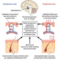
Regulation: Maintains body temperature, pH, and fluid volume in the circulatory system.
Protection: Prevents blood loss via clotting and defends against infection through immune cells and proteins.
Composition and Physical Characteristics of Blood
Physical Properties
Blood is a sticky, opaque fluid with a metallic taste.
Temperature: 38ºC (100.4ºF)
Viscosity: High
pH: Slightly alkaline (7.35–7.45)
Volume: ~8% of body weight; males 5–6 L, females 4–5 L
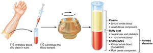
Components of Blood
Blood consists of plasma (fluid matrix) and formed elements (cells and cell fragments).
Plasma: Straw-colored, sticky fluid, about 90% water, contains over 100 dissolved solutes.
Formed Elements: Erythrocytes (RBCs), leukocytes (WBCs), and platelets.
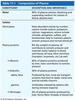
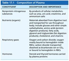
Blood Plasma: Composition
Plasma is the nonliving fluid component of blood, containing water, proteins, electrolytes, nutrients, gases, hormones, and waste products.
Albumin: Most abundant plasma protein; maintains osmotic pressure and acts as a carrier.
Globulins: Transport proteins and antibodies.
Fibrinogen: Forms fibrin threads for blood clotting.
Constituent | Description and Importance |
|---|---|
Water | 90% of plasma; dissolving and suspending medium for solutes |
Electrolytes | Maintain osmotic balance, pH, and membrane potential |
Plasma proteins | Albumin, globulins, fibrinogen; osmotic pressure, transport, immunity, clotting |
Constituent | Description and Importance |
|---|---|
Nonprotein nitrogenous substances | By-products of metabolism (urea, uric acid, creatinine) |
Nutrients | Glucose, amino acids, fatty acids, vitamins |
Respiratory gases | Oxygen and carbon dioxide |
Hormones | Steroid and thyroid hormones carried by plasma proteins |
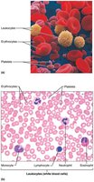
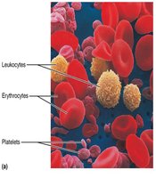
Formed Elements of Blood
Types of Formed Elements
The formed elements include erythrocytes, leukocytes, and platelets.
Erythrocytes (RBCs): Transport oxygen and carbon dioxide.
Leukocytes (WBCs): Defend against infection.
Platelets: Cell fragments involved in clotting.
Erythrocytes (Red Blood Cells)
Structure and Function
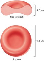
Erythrocytes are small, biconcave, anucleate cells specialized for gas transport.
Biconcave shape increases surface area for gas exchange.
Filled with hemoglobin (Hb), which binds oxygen.
No mitochondria; ATP production is anaerobic.
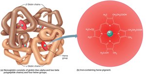
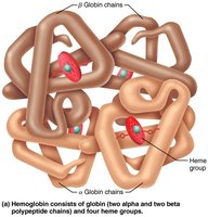
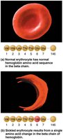
Hemoglobin Structure and Function
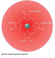
Hemoglobin is a protein composed of four polypeptide chains (two alpha, two beta), each with a heme group containing iron.
Each heme binds one O2 molecule.
Each RBC contains ~250 million Hb molecules.
Hemoglobin also binds CO2 (carbaminohemoglobin).
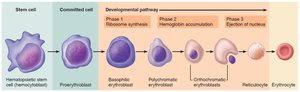
Erythropoiesis: Formation of Red Blood Cells
Erythropoiesis is the process of RBC formation, occurring in red bone marrow.
Stem cell (hemocytoblast) differentiates into myeloid stem cell, then proerythroblast, and through several stages to mature erythrocyte.
Reticulocyte count indicates rate of RBC formation.
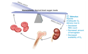
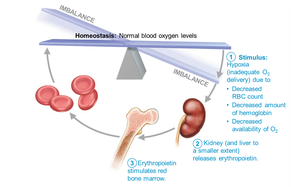
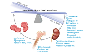
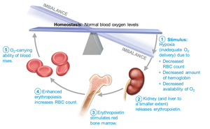
Regulation of Erythropoiesis
Erythropoiesis is regulated by erythropoietin (EPO), a hormone released by kidneys in response to hypoxia (low O2).
Balance between RBC production and destruction is critical.
Dietary requirements include amino acids, iron, vitamin B12, and folic acid.


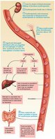
Fate and Destruction of Erythrocytes
RBCs have a lifespan of 100–120 days. Old RBCs are destroyed by macrophages in the spleen, and their components are recycled.
Heme is degraded to bilirubin, excreted in bile.
Iron is stored or reused.
Globin is broken down to amino acids.
Erythrocyte Disorders
Anemia
Anemia is a condition of reduced O2-carrying capacity. Causes include blood loss, insufficient RBC production, or excessive RBC destruction.
Iron-deficiency anemia: Microcytic, hypochromic RBCs; treated with iron supplements.
Pernicious anemia: Lack of intrinsic factor for B12 absorption; treated with B12 injections.
Renal anemia: Lack of EPO; treated with synthetic EPO.
Aplastic anemia: Bone marrow destruction; treated with transfusions or stem cell transplants.
Hemolytic anemias: Premature RBC lysis; includes thalassemias and sickle-cell anemia.
Polycythemia
Polycythemia is an abnormal excess of RBCs, increasing blood viscosity.
Polycythemia vera: Bone marrow cancer.
Secondary polycythemia: Due to low O2 or increased EPO.
Blood doping: Artificially increasing RBC count for athletic performance.
Leukocytes (White Blood Cells)
Structure and Function
Leukocytes are complete cells with nuclei and organelles, functioning in defense against disease.
Can leave capillaries (diapedesis) and move through tissues (amoeboid motion).
Leukocytosis: Elevated WBC count, normal response to infection.
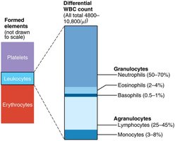

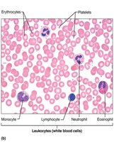
Types of Leukocytes
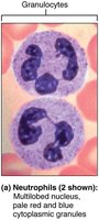
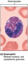
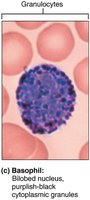
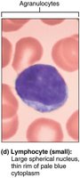
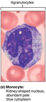
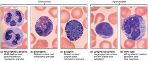
Leukocytes are classified as granulocytes (neutrophils, eosinophils, basophils) and agranulocytes (lymphocytes, monocytes).
Neutrophils: Most numerous; phagocytic; "bacteria slayers".
Eosinophils: Digest parasitic worms; modulate immune response.
Basophils: Release histamine; rarest WBC.
Lymphocytes: Crucial to immunity; T cells and B cells.
Monocytes: Largest WBC; become macrophages in tissues.
Production and Life Span of Leukocytes
Leukopoiesis
Leukopoiesis is the formation of WBCs, stimulated by interleukins and colony-stimulating factors (CSFs).
All leukocytes originate from hemocytoblasts.
Lymphoid stem cells produce lymphocytes; myeloid stem cells produce other WBCs.
Platelets
Structure and Function
Platelets are fragments of megakaryocytes, essential for blood clotting.
Contain chemicals for clotting (serotonin, calcium, enzymes, ADP).
Form temporary plugs to seal vessel breaks.
Hemostasis
Steps of Hemostasis
Hemostasis is the process of stopping bleeding, involving three steps:
Vascular spasm: Vasoconstriction in response to injury.
Platelet plug formation: Platelets adhere to exposed collagen and release chemicals to attract more platelets.
Coagulation: Reinforces plug with fibrin threads; involves intrinsic and extrinsic pathways leading to thrombin and fibrin formation.
Blood Transfusions and Blood Typing
Blood Groups
Blood groups are determined by antigens (agglutinogens) on RBC membranes.
ABO blood groups: Based on presence of A and B antigens.
Rh blood group: Presence or absence of Rh antigen (D).
Transfusion Reactions
Mismatched transfusions can cause agglutination and destruction of donor RBCs, leading to serious complications.
Diagnostic Blood Tests
Common Tests
Hematocrit: Measures RBC proportion.
Blood glucose: Checks for diabetes.
Differential WBC count: Assesses immune status.
Complete blood count (CBC): Evaluates overall blood health.
Developmental Aspects of Blood
Fetal and Aging Blood
Fetal blood cells form in yolk sac, liver, and spleen; red bone marrow becomes primary site by seventh month.
Hemoglobin F in fetus has higher O2 affinity than adult hemoglobin A.
Blood diseases of aging include chronic leukemias, anemias, and clotting disorders.
Key Equations and Concepts
Hematocrit Calculation
Hematocrit (%) = (Volume of RBCs / Total blood volume) × 100
Oxygen Transport by Hemoglobin
Each hemoglobin molecule can bind four O2 molecules.
Blood Volume Estimation
Blood volume (L) = 0.07 × body weight (kg)
Coagulation Pathways
Intrinsic and extrinsic pathways converge at factor X activation, leading to thrombin and fibrin formation.
Sample Equation (Hemoglobin-Oxygen Binding)
Sample Equation (Hematocrit)
Sample Equation (Blood Volume)
Sample Equation (Coagulation Pathway)
Summary Table: Formed Elements of Blood
Element | Main Function | Key Features |
|---|---|---|
Erythrocytes | O2 and CO2 transport | Biconcave, anucleate, hemoglobin-rich |
Leukocytes | Defense against infection | Complete cells, various types |
Platelets | Clotting | Cell fragments, contain clotting factors |
Summary Table: Leukocyte Types
Type | Relative Abundance | Main Function |
|---|---|---|
Neutrophils | 50–70% | Phagocytosis of bacteria |
Lymphocytes | 25–45% | Immunity (T and B cells) |
Monocytes | 3–8% | Phagocytosis, become macrophages |
Eosinophils | 2–4% | Digest parasitic worms, modulate immunity |
Basophils | 0.5–1% | Release histamine, inflammation |
Summary Table: ABO Blood Groups
Blood Type | Antigens Present | Antibodies Present |
|---|---|---|
A | A | Anti-B |
B | B | Anti-A |
AB | A and B | None |
O | None | Anti-A and Anti-B |
