 Back
BackStudy Notes: The Nervous System – Structure, Function, and Protection
Study Guide - Smart Notes
 Tailored notes based on your materials, expanded with key definitions, examples, and context.
Protection of the Central Nervous System (CNS)
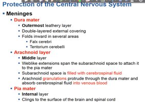
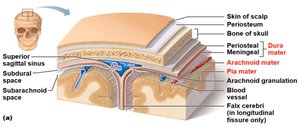
Meninges
The meninges are three connective tissue membranes that cover and protect the brain and spinal cord. They provide structural support, contain cerebrospinal fluid, and help form a barrier against pathogens.
Dura mater: The outermost, tough, double-layered membrane. It forms inward folds in certain areas (e.g., falx cerebri, tentorium cerebelli).
Arachnoid mater: The middle, web-like membrane. The subarachnoid space beneath it is filled with cerebrospinal fluid (CSF). Arachnoid granulations absorb CSF into venous blood.
Pia mater: The innermost, delicate membrane that clings tightly to the surface of the brain and spinal cord.
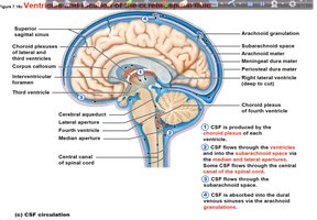
Cerebrospinal Fluid (CSF)
Cerebrospinal fluid is a clear, colorless liquid that surrounds the brain and spinal cord, providing cushioning, buoyancy, and chemical stability. It is produced by the choroid plexuses in the ventricles of the brain and circulates through the ventricular system and subarachnoid space.
CSF protects the CNS from trauma and removes metabolic waste.
It is absorbed into the venous blood via arachnoid granulations.
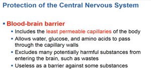
Blood-Brain Barrier
The blood-brain barrier is a selective permeability barrier formed by endothelial cells of CNS capillaries. It protects the brain from harmful substances while allowing essential nutrients to pass through.
Composed of the least permeable capillaries in the body.
Permits water, glucose, and amino acids; restricts toxins and pathogens.
Not effective against some substances (e.g., lipid-soluble molecules, certain drugs).
Spinal Cord
Structure and Function
The spinal cord extends from the foramen magnum of the skull to the first or second lumbar vertebra. It serves as a two-way conduction pathway between the brain and the body and is the site of spinal reflexes.
31 pairs of spinal nerves arise from the spinal cord.
The cauda equina is a bundle of spinal nerves at the inferior end.
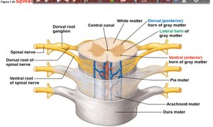
Cross-Sectional Anatomy
The spinal cord consists of central gray matter (containing neuron cell bodies) and surrounding white matter (containing myelinated axons). The meninges surround the cord, providing protection.
Dorsal (posterior) and ventral (anterior) horns of gray matter are present.
White matter is organized into columns carrying ascending and descending tracts.
Peripheral Nervous System (PNS)
Overview
The peripheral nervous system consists of nerves and ganglia outside the CNS. It connects the CNS to limbs and organs, facilitating communication throughout the body.
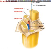
Structure of a Nerve
Nerves are bundles of neuron fibers (axons) found outside the CNS. Each nerve fiber is surrounded by connective tissue layers:
Endoneurium: Surrounds individual nerve fibers.
Perineurium: Wraps groups of fibers into fascicles.
Epineurium: Binds groups of fascicles together to form the nerve.
Types of Nerves
Mixed nerves: Contain both sensory (afferent) and motor (efferent) fibers.
Sensory (afferent) nerves: Carry impulses toward the CNS.
Motor (efferent) nerves: Carry impulses away from the CNS.
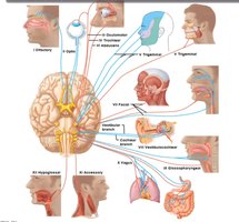
Cranial Nerves
There are 12 pairs of cranial nerves, most of which serve the head and neck. The vagus nerves are the only pair that extends into the thoracic and abdominal cavities. Most cranial nerves are mixed, but three are purely sensory: optic, olfactory, and vestibulocochlear.
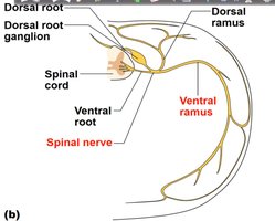
Spinal Nerves
There are 31 pairs of spinal nerves, each formed by the combination of ventral and dorsal roots of the spinal cord. They are named according to the region of the spinal cord from which they arise.
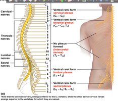
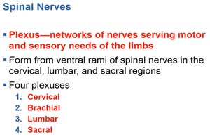
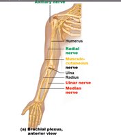
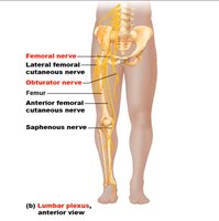
Plexuses
Plexuses are networks of nerves serving the motor and sensory needs of the limbs. They are formed from the ventral rami of spinal nerves in the cervical, lumbar, and sacral regions. The four main plexuses are cervical, brachial, lumbar, and sacral.
Autonomic Nervous System (ANS)
Overview
The autonomic nervous system is a motor subdivision of the PNS that controls involuntary body functions. It regulates cardiac and smooth muscle activity, as well as glandular function.
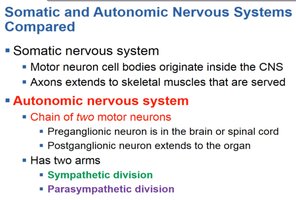
Somatic vs. Autonomic Nervous Systems
Somatic nervous system: Motor neuron cell bodies originate in the CNS; axons extend to skeletal muscles.
Autonomic nervous system: Involves a chain of two motor neurons (preganglionic and postganglionic). Has two divisions: sympathetic and parasympathetic.
Sympathetic Division
The sympathetic division (thoracolumbar division) prepares the body for "fight or flight" responses. Preganglionic neurons originate from T1 to L2 spinal segments. It increases heart rate, dilates bronchioles, and inhibits digestion during stress or emergency.
Axons pass through a ramus communicans to enter a sympathetic trunk ganglion.
Sympathetic trunk lies near the spinal cord.
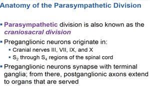
Parasympathetic Division
The parasympathetic division (craniosacral division) promotes "rest-and-digest" activities. Preganglionic neurons originate in cranial nerves III, VII, IX, X, and S2–S4 spinal segments. It conserves energy and maintains daily body functions.
Autonomic Functioning
Sympathetic division: "E" division—exercise, excitement, emergency, embarrassment.
Parasympathetic division: "D" division—digestion, defecation, diuresis.
Summary Table: Comparison of Somatic and Autonomic Nervous Systems
Feature | Somatic Nervous System | Autonomic Nervous System |
|---|---|---|
Effectors | Skeletal muscle | Cardiac muscle, smooth muscle, glands |
Number of neurons in pathway | One | Two (preganglionic and postganglionic) |
Neurotransmitter(s) | Acetylcholine | Acetylcholine, norepinephrine |
Control | Voluntary | Involuntary |
