 Back
BackThe Autonomic Nervous System: Structure, Function, and Comparison with the Somatic Nervous System
Study Guide - Smart Notes
 Tailored notes based on your materials, expanded with key definitions, examples, and context.
The Autonomic Nervous System (ANS)
Introduction to the Autonomic Nervous System
The autonomic nervous system (ANS) is a division of the peripheral nervous system responsible for regulating involuntary physiological processes, including heart rate, blood pressure, respiration, digestion, and sexual arousal. It operates largely below the level of consciousness and controls visceral functions by innervating smooth muscle, cardiac muscle, and glands.
Main Divisions: Sympathetic and Parasympathetic Nervous Systems
Primary Function: Maintain homeostasis by balancing bodily responses to internal and external stimuli
Review Questions: Cranial Nerves and Spinal Cord Organization
Cranial Nerves and Hearing
The cranial nerve responsible for transducing hearing is Cranial Nerve VIII, also known as the Vestibulocochlear Nerve. This nerve carries auditory sensory information from the cochlea of the inner ear directly to the brain.
Other Cranial Nerves: The Glossopharyngeal Nerve (IX), Cranial Nerve VI (Abducens), and the Vagus Nerve (X) do not transduce hearing.
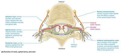
Sensory Neurons and the Spinal Cord
Sensory neurons enter the spinal cord at the Posterior Root Ganglion. This structure contains the cell bodies of sensory neurons and is a critical relay point for sensory information entering the central nervous system.
Posterior Horn: Receives sensory information from the posterior root ganglion.
Anterior Horn: Contains motor neurons that send signals to skeletal muscles.
Anterior Ramus: Carries both sensory and motor fibers to and from the limbs and anterior trunk.
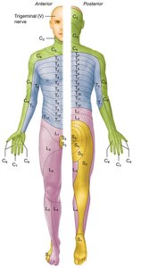
Dermatomes
Dermatomes are regions of the skin innervated by sensory fibers from a single spinal nerve. Mapping dermatomes is clinically important for diagnosing nerve or spinal cord injuries.
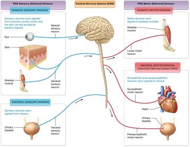
Organization of the Peripheral Nervous System (PNS)
Overview of PNS Structure
The PNS is divided into sensory (afferent) and motor (efferent) divisions. The motor division is further subdivided into the somatic nervous system (controls voluntary movements) and the autonomic nervous system (controls involuntary functions).
Somatic Sensory Division: Transmits sensory information from skin, muscles, and joints to the CNS.
Visceral Sensory Division: Transmits sensory information from organs to the CNS.
Somatic Motor Division: Controls voluntary movements via skeletal muscles.
Autonomic Motor Division: Controls involuntary responses via smooth muscle, cardiac muscle, and glands.
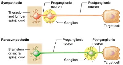
Somatic vs. Autonomic Nervous Systems
Comparison of Structure and Function
The somatic and autonomic nervous systems differ in their target tissues, control mechanisms, and neural pathways.
Somatic Nervous System: Single neuron pathway from CNS to skeletal muscle; voluntary control.
Autonomic Nervous System: Two-neuron pathway (preganglionic and postganglionic) from CNS to target organ; involuntary control.
Visceral Reflex Arcs
Mechanism of Visceral Reflexes
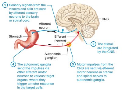
Visceral reflex arcs are the basic functional units of the ANS, allowing for automatic responses to changes in the internal environment. These reflexes involve sensory input from visceral organs, integration in the CNS, and motor output to effectors via autonomic ganglia.
Sensory signals from viscera and skin are sent by afferent sensory neurons to the brain or spinal cord.
The CNS integrates the stimuli.
Motor impulses from the CNS are sent via efferent motor neurons to autonomic ganglia.
The autonomic ganglia send impulses via other efferent motor neurons to target organs, triggering a response.
Divisions of the Autonomic Nervous System
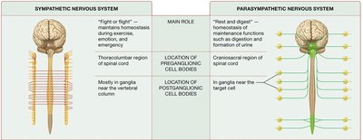
Sympathetic Nervous System (Thoracolumbar Division)
The sympathetic nervous system prepares the body for 'fight or flight' responses. Preganglionic neurons originate in the thoracic and lumbar spinal cord and synapse in sympathetic ganglia near the spinal cord.
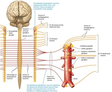
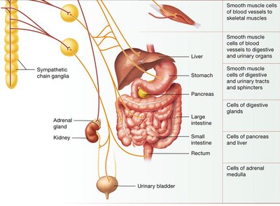
Sympathetic Chain Ganglia: Series of ganglia adjacent to the vertebral column where most preganglionic neurons synapse.
Collateral Ganglia: Ganglia located anterior to the vertebral column, involved in innervating abdominal and pelvic organs.
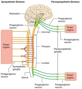
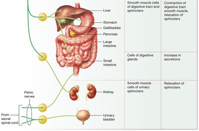
Parasympathetic Nervous System (Craniosacral Division)
The parasympathetic nervous system supports 'rest and digest' activities. Preganglionic neurons originate in the brainstem and sacral spinal cord and synapse in ganglia near or within target organs (terminal ganglia).
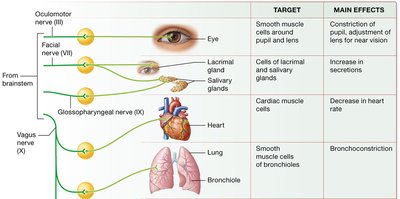
Major Cranial Nerves: Vagus nerve (X), Oculomotor (III), Facial (VII), Glossopharyngeal (IX)
Function: Promotes digestion, energy storage, and maintenance functions.
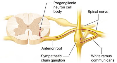
Sympathetic Chain Ganglia and White Ramus Communicans
Pathways of Sympathetic Neurons
All preganglionic sympathetic neurons must pass through the white ramus communicans to reach the sympathetic chain ganglia. From there, they may synapse within the chain or pass through to collateral ganglia.
White Ramus Communicans: Pathway for preganglionic fibers entering the sympathetic chain.
Sympathetic Chain Ganglia: Relay points for sympathetic signals to various organs.
Neurotransmitters and Receptors in the ANS
Sympathetic Neurotransmitters
Sympathetic preganglionic neurons release acetylcholine (ACh) at synapses with postganglionic neurons. Postganglionic neurons typically release norepinephrine (NE) or epinephrine at target organs, which bind to adrenergic receptors (alpha or beta types). Some postganglionic neurons release ACh, especially those innervating sweat glands.
Adrenergic Receptors: Bind NE or epinephrine; divided into alpha and beta subtypes.
Cholinergic Receptors: Bind ACh; divided into nicotinic (on postganglionic neurons) and muscarinic (on target cells) types.
Parasympathetic Neurotransmitters
Both preganglionic and postganglionic parasympathetic neurons release acetylcholine (ACh). All postganglionic neurons have nicotinic receptors, while all parasympathetic target cells have muscarinic receptors.
Effects of the Sympathetic and Parasympathetic Nervous Systems
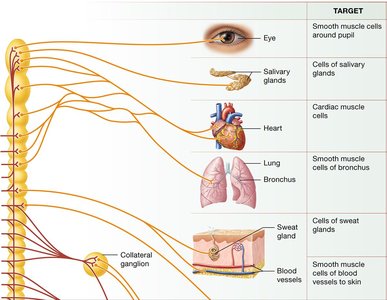
Sympathetic Nervous System Targets and Effects
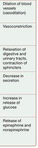
The sympathetic nervous system innervates a wide range of organs, producing effects that prepare the body for rapid action.
Target | Main Effects |
|---|---|
Eye (pupil) | Dilation of pupil |
Salivary glands | Increase in secretion in certain cells |
Heart | Increase in heart rate and force of contraction |
Lung (bronchioles) | Dilation of bronchioles (bronchodilation) |
Sweat glands | Increase in secretion |
Blood vessels to skin | Constriction of blood vessels (vasoconstriction) |
Liver | Increase in release of glucose |
Adrenal medulla | Release of epinephrine and norepinephrine |
Digestive tract | Relaxation of digestive and urinary tracts, contraction of sphincters, decrease in secretion |
Parasympathetic Nervous System Targets and Effects
The parasympathetic nervous system generally has effects that conserve energy and promote maintenance activities.
Nerve | Target | Main Effects |
|---|---|---|
Oculomotor (III) | Eye | Constriction of pupil, adjustment of lens for near vision |
Facial (VII) | Lacrimal and salivary glands | Increase in secretions |
Glossopharyngeal (IX) | Salivary glands | Increase in secretions |
Vagus (X) | Heart | Decrease in heart rate |
Vagus (X) | Lung (bronchioles) | Bronchoconstriction |
Vagus (X) | Digestive tract | Contraction of digestive tract smooth muscle, relaxation of sphincters, increase in secretion |
Pelvic nerves | Urinary bladder | Relaxation of sphincters |
Comparison of the Sympathetic and Parasympathetic Nervous Systems
Summary Table
Sympathetic Nervous System | Parasympathetic Nervous System | |
|---|---|---|
Main Role | "Fight or flight" – maintains homeostasis during exercise, excitement, and emergency | "Rest and digest" – maintenance functions such as digestion and formation of urine |
Location of Preganglionic Cell Bodies | Thoracolumbar region of spinal cord | Craniosacral region of spinal cord |
Location of Postganglionic Cell Bodies | Mostly in ganglia near the vertebral column | In ganglia near the target cell |
Neurotransmitter and Receptor Comparison Table
Sympathetic | Parasympathetic | |
|---|---|---|
PREGANGLIONIC NEUROTRANSMITTERS | Acetylcholine (ACh) | Acetylcholine (ACh) |
POSTGANGLIONIC RECEPTORS | Nicotinic | Nicotinic |
POSTGANGLIONIC NEUROTRANSMITTERS | Norepinephrine (NE), Epinephrine (Epi), or ACh | Acetylcholine (ACh) |
TARGET CELL RECEPTORS | Adrenergic (alpha, beta), Muscarinic (for ACh) | Muscarinic |
Summary
The autonomic nervous system is essential for maintaining homeostasis and responding to changes in the internal and external environment. Understanding the structure, function, and neurotransmitter mechanisms of the sympathetic and parasympathetic divisions is crucial for comprehending how the body regulates involuntary processes.
