 Back
BackThe Cardiovascular System: Blood and Blood Vessels
Study Guide - Smart Notes
 Tailored notes based on your materials, expanded with key definitions, examples, and context.
The Cardiovascular System: Overview
Major Components and Functions
The cardiovascular system is a closed circulatory system consisting of the heart, blood vessels, and blood. Its primary function is to transport nutrients, gases, hormones, and waste products throughout the body.
Blood: A connective tissue composed of plasma and formed elements (cells).
Blood Vessels: Arteries, veins, and capillaries that deliver blood to all tissues.
Heart: A muscular pump that moves blood through the vessels.
General Functions
Transport: Oxygen, nutrients, hormones, and waste products.
Defense: White blood cells and antibodies protect against pathogens.
Regulation: Osmosis, pH, and temperature.
Hemostasis: Platelets and clotting proteins minimize blood loss.
Blood: Composition and Properties
Physical Characteristics
Blood is a liquid tissue, slightly more viscous than water, with a metallic, salty taste. Its color varies from bright red (oxygenated) to purplish (deoxygenated).
Temperature: 37°C
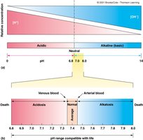
pH: 7.35–7.45 (slightly alkaline)
Volume: 6–8% of body weight (4–6 quarts in adults)
Blood pH and Life Compatibility
Blood pH is tightly regulated.
Acidosis: pH < 7.35
Alkalosis: pH > 7.45
Extreme pH (<6.8 or >8.0) is incompatible with life.
Blood Components
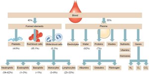
Blood separates into plasma (liquid) and formed elements (cells) when centrifuged.
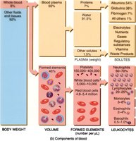
Plasma: 50–60% of blood volume, mostly water, contains nutrients, waste, proteins, and electrolytes.
Formed Elements: Erythrocytes (RBCs), leukocytes (WBCs), and platelets.
Major Classes of Blood Proteins
Albumins: Maintain osmotic pressure and transport hydrophobic molecules.
Globulins: Antibodies for immune response.
Fibrinogen: Blood clotting factor.
Formed Elements of Blood
Erythrocytes (Red Blood Cells)
Erythrocytes are biconcave, flexible, and lack nuclei in mammals. Their main function is gas transport.
Hemoglobin: Iron-containing protein that binds O2 and CO2.
Hematocrit: Percentage of blood volume made up of RBCs (males: 40–54%, females: 37–47%).
Leukocytes (White Blood Cells)
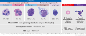
Leukocytes are involved in defense and housekeeping.
Phagocytes: Neutrophils, monocytes, macrophages (engulf foreign particles).
Immunocytes: Lymphocytes (specific immune responses).
Granulocytes: Neutrophils, eosinophils, basophils (contain granules).
Thrombocytes (Platelets)
Platelets are cell fragments involved in clotting.
Non-nucleated, membrane-bound fragments of megakaryocytes.
Hemopoiesis: Blood Cell Formation
Hematopoietic Stem Cells
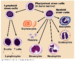
Blood cell production begins with stem cells in the bone marrow.
Myeloid Stem Cells: Form erythrocytes, platelets, granulocytes, monocytes.
Lymphoid Stem Cells: Form lymphocytes (B, T, and NK cells).
Erythropoiesis
Erythrocyte formation is regulated by erythropoietin (EPO), released in response to low oxygen.
Stem cells → erythroblasts → reticulocytes → erythrocytes
Reticulocytes mature into erythrocytes within 24 hours.
RBC lifespan: ~120 days
Erythrocyte Recycling and Jaundice
RBC Breakdown and Recycling
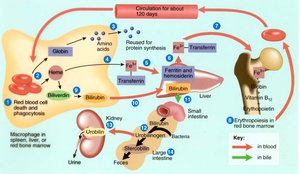
After ~120 days, RBCs are removed by the spleen and broken down by macrophages.
Hemoglobin is split into globin (protein) and heme (iron-containing pigment).
Iron is reused; heme is converted to bilirubin.
Bilirubin is carried to the liver, used to make bile, and excreted in urine and feces.
Jaundice
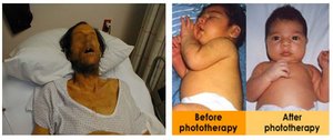
Elevated bilirubin levels (hyperbilirubinemia) cause jaundice, resulting in yellowing of skin and eyes.
Caused by liver disease or rapid breakdown of fetal hemoglobin.
Treated with phototherapy in newborns.
Blood Vessels: Structure and Function
Layers of Blood Vessel Walls
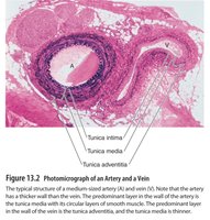
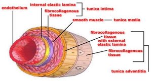
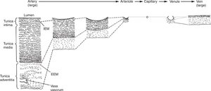
Most blood vessels (except capillaries) have three layers (tunics):
Tunica intima: Innermost layer, simple squamous epithelium, internal elastic lamina.
Tunica media: Middle layer, smooth muscle, controls vasoconstriction and dilation.
Tunica adventitia: Outermost layer, fibrocollagenous tissue, anchors vessels.
Arteries vs. Veins
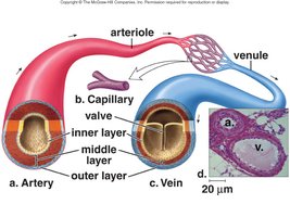
Arteries: Thick, muscular, elastic walls; function as resistance vessels; carry blood away from the heart.
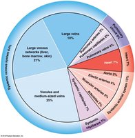
Veins: Thinner, more compliant walls; function as blood reservoirs; carry blood toward the heart.
Capillaries: Types and Functions
Capillary Beds and Exchange
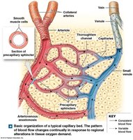
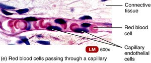
Capillaries are the site of gas, nutrient, and waste exchange.
Walls are one cell thick (endothelium only).
Capillary beds are regulated by precapillary sphincters.
Density correlates with tissue metabolic activity.
Types of Capillaries
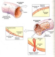
Continuous: No holes; most common; found in muscle, connective tissue, nervous tissue.
Fenestrated: Small pores; found in kidneys and small intestine.
Sinusoids (Discontinuous): Large gaps; found in liver and bone marrow.
Venous Return and Varicose Veins
Mechanisms of Venous Return
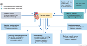
Venous return is aided by several mechanisms:
Sympathetic activity: Increases venous tone.
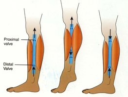
Skeletal muscle pump: Muscle contractions squeeze veins, pushing blood toward the heart.
Venous valves: Prevent backflow.
Respiratory pump: Changes in thoracic pressure during breathing aid blood flow.
Cardiac suction: Negative pressure in atria during ventricular emptying enhances return.
Varicose Veins
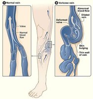
Varicose veins are dilated, twisted veins caused by leaky valves, leading to blood pooling.
Common in legs and around the anus (hemorrhoids).
Can be congenital or due to physical stress.
Hemostasis: Minimizing Blood Loss
Three Main Methods
Vascular Spasm: Local vasoconstriction reduces blood flow.
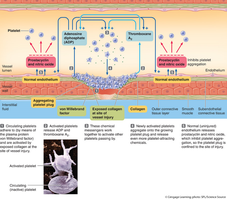
Platelet Plug: Platelets adhere to exposed collagen, aggregate, and form a plug.
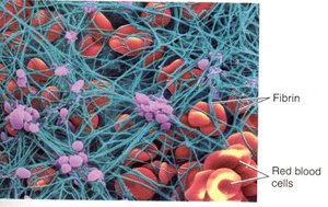
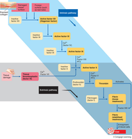
Clot Formation: Coagulation cascade activates thrombin, which converts fibrinogen to fibrin, forming a meshwork that traps blood cells.
Clot Retraction and Dissolution
Platelets contract, shrinking the clot and pulling vessel edges together.
Fibroblasts form a scar at the defect.
Plasmin dissolves fibrin, removing the clot.
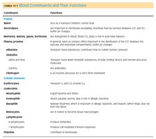
Summary Table: Blood Constituents and Functions
Constituent | Function |
|---|---|
Water | Acts as a transport medium; carries heat |
Electrolytes | Maintain osmotic balance; buffer pH changes |
Proteins | Osmotic pressure, transport, immune response, clotting |
Albumins | Osmotic pressure, carrier for hydrophobic molecules |
Globulins | Antibodies, transport proteins |
Fibrinogen | Clotting factor |
Erythrocytes | Transport O2 and CO2 |
Leukocytes | Defense and housekeeping |
Platelets | Clotting |
Key Equations
Osmotic Pressure
Where is osmotic pressure, is the van't Hoff factor, is molarity, is the gas constant, and is temperature.
pH Calculation
Where is the concentration of hydrogen ions.
Hemoglobin Oxygen Binding
Hemoglobin binds up to four oxygen molecules.
Coagulation Cascade
Thrombin converts fibrinogen to fibrin, forming the clot.
Additional info:
Blood vessel structure determines function: arteries are resistance vessels due to thick muscular walls; veins are compliance vessels due to thinner, elastic walls.
Capillary density is highest in tissues with high metabolic activity.
Feedback mechanisms regulate platelet aggregation to prevent excessive clot formation.
