 Back
BackThe Cardiovascular System: Structure and Function
Study Guide - Smart Notes
 Tailored notes based on your materials, expanded with key definitions, examples, and context.
The Cardiovascular System
Overview and Functions
The cardiovascular system is a closed system composed of the heart and blood vessels. Its primary function is to transport oxygen, nutrients, cell wastes, and hormones to and from the cells of the body. The heart acts as a pump, while blood vessels provide the route for blood circulation throughout the body.
Transport: Delivers essential substances to tissues and removes metabolic wastes.
Regulation: Maintains homeostasis, including temperature and pH balance.
Protection: Circulates immune cells and clotting factors.
Anatomy of the Heart
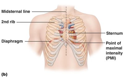
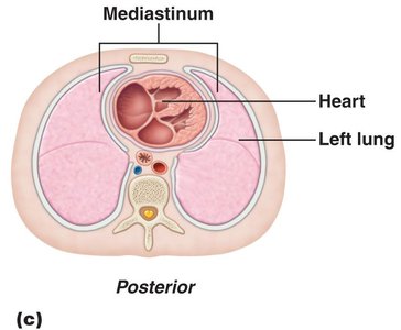
Location and Orientation
The heart is approximately the size of a human fist and weighs less than a pound. It is located in the thoracic cavity, between the lungs in the inferior mediastinum. The apex points toward the left hip and rests on the diaphragm, while the base points toward the right shoulder.
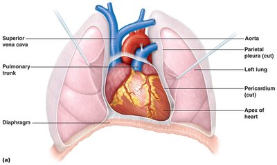
Heart Wall and Coverings
The heart wall consists of three layers, each with distinct functions and structures. The heart is also enclosed by a double-walled sac called the pericardium, which protects and anchors the heart.
Fibrous Pericardium: Tough, dense connective tissue that anchors the heart and prevents overexpansion.
Serous Pericardium: Composed of a parietal layer (lining the fibrous pericardium) and a visceral layer (epicardium) that covers the heart surface.

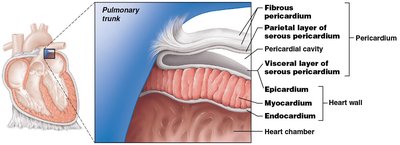
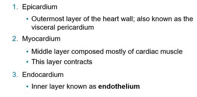
Epicardium: Outermost layer; also known as the visceral pericardium.
Myocardium: Middle layer composed of cardiac muscle; responsible for contraction.
Endocardium: Inner layer; a thin endothelium lining the heart chambers.
Clinical Note: Pericarditis
Pericarditis: Inflammation of the pericardium, often caused by infection. Can lead to fluid accumulation (cardiac tamponade), restricting heart movement and reducing cardiac output.
Chambers and Associated Great Vessels
Heart Chambers
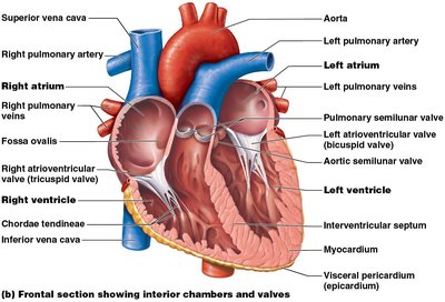
The heart contains four chambers: two atria (superior, receiving chambers) and two ventricles (inferior, discharging chambers). The atria receive blood under low pressure from veins, while the ventricles have thick walls and pump blood into circulation.
Septa of the Heart
Interatrial septum: Separates the right and left atria.
Interventricular septum: Separates the right and left ventricles.

Double Pump Function
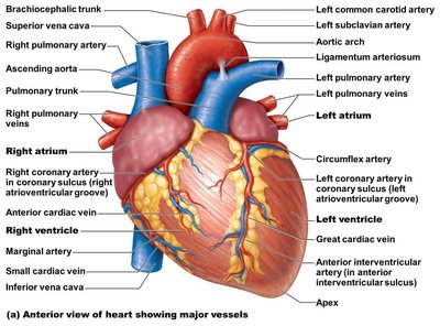
Pulmonary Circuit: Right side pumps deoxygenated blood to the lungs via the pulmonary trunk and arteries; oxygenated blood returns via pulmonary veins to the left atrium.
Systemic Circuit: Left side pumps oxygenated blood to the body via the aorta; deoxygenated blood returns to the right atrium via the superior and inferior vena cava.

Heart Valves
Types and Functions
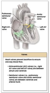
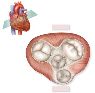
Heart valves ensure unidirectional blood flow and prevent backflow. There are two main types:
Atrioventricular (AV) Valves: Between atria and ventricles (right: tricuspid; left: bicuspid/mitral).
Semilunar Valves: Between ventricles and major arteries (pulmonary and aortic semilunar valves).
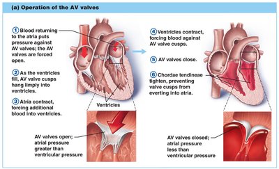
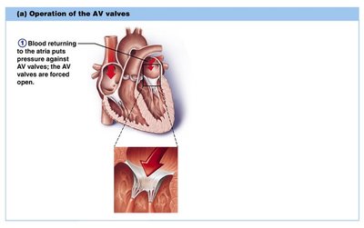
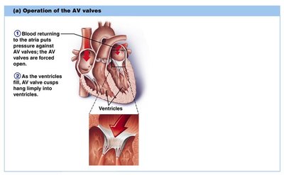
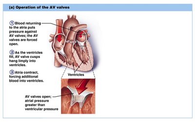
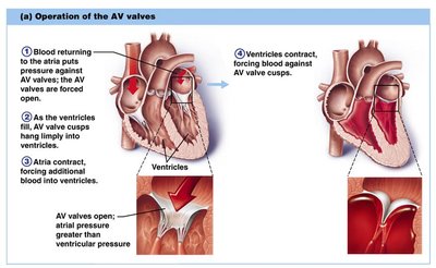
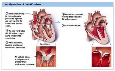
Valve Operation
AV Valves: Anchored by chordae tendineae; open during relaxation, closed during contraction.
Semilunar Valves: Closed during relaxation, open during ventricular contraction.
Valves open and close in response to pressure changes within the heart.
Heart Wall: Fibrous Skeleton and Contraction Pattern
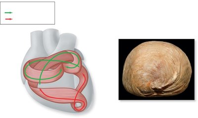
Fibrous Skeleton
The fibrous skeleton of the heart provides structural support and anchors cardiac muscle fibers. Cardiac muscle cells are arranged in spiral bundles, allowing efficient contraction patterns.
Atrial contraction: Compresses chamber walls to move blood into ventricles.
Ventricular contraction: Begins at the apex and compresses superiorly, similar to wringing a mop.
Summary Table: Heart Chambers and Valves
Chamber | Receives Blood From | Pumps Blood To | Valve |
|---|---|---|---|
Right Atrium | Superior/Inferior Vena Cava, Coronary Sinus | Right Ventricle | Tricuspid (Right AV) |
Right Ventricle | Right Atrium | Pulmonary Trunk | Pulmonary Semilunar |
Left Atrium | Pulmonary Veins | Left Ventricle | Bicuspid (Mitral, Left AV) |
Left Ventricle | Left Atrium | Aorta | Aortic Semilunar |
Key Terms and Concepts
Pericardium: Double-walled sac enclosing the heart.
Epicardium: Outermost layer of the heart wall.
Myocardium: Muscular, contractile middle layer of the heart wall.
Endocardium: Thin, endothelial inner lining of the heart chambers.
Chordae Tendineae: Tendinous cords anchoring AV valve cusps to ventricular walls.
Fibrous Skeleton: Dense connective tissue framework supporting the heart.
Additional info: The heart's double pump function is essential for separating pulmonary and systemic circulation, ensuring efficient oxygenation of blood and delivery to tissues. The fibrous skeleton also acts as an electrical insulator, preventing direct transmission of electrical impulses between atria and ventricles.
