 Back
BackThe Cardiovascular System: Structure and Function of the Heart, Arteries, and Veins
Study Guide - Smart Notes
 Tailored notes based on your materials, expanded with key definitions, examples, and context.
The Cardiovascular System
Overview of Blood Circulation
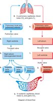
The cardiovascular system is responsible for the transport of blood throughout the body, delivering oxygen and nutrients while removing waste products. Blood circulates through two main circuits: the pulmonary circuit (to and from the lungs) and the systemic circuit (to and from the rest of the body).
Pulmonary Circuit: Carries deoxygenated blood from the right side of the heart to the lungs and returns oxygenated blood to the left side of the heart.
Systemic Circuit: Distributes oxygenated blood from the left side of the heart to the body and returns deoxygenated blood to the right side of the heart.
Key Structures: Heart chambers, valves, arteries, veins, capillaries.
Blood Flow: Blue indicates deoxygenated blood; red indicates oxygenated blood.
Example: The right atrium receives deoxygenated blood from the body, which is then pumped to the lungs via the pulmonary artery.
Structure of the Heart
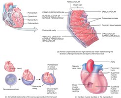
The heart is a muscular organ enclosed by the pericardium, which protects and anchors it. The heart wall consists of three layers: endocardium, myocardium, and epicardium. The pericardium itself has two main components: the fibrous pericardium and the serous pericardium.
Fibrous Pericardium: Dense irregular connective tissue that protects, anchors, and prevents overstretching of the heart.
Serous Pericardium: Thin, delicate membrane with a parietal layer (outer), pericardial cavity (fluid-filled), and visceral layer (epicardium).
Heart Wall Layers:
Endocardium: Inner lining of the heart chambers.
Myocardium: Thick middle layer of cardiac muscle responsible for contraction.
Epicardium: Outer layer, also the visceral layer of the serous pericardium.
Example: The pericardial fluid reduces friction between the heart and surrounding structures during contraction.
Heart Chambers and Valves
The heart contains four chambers: right atrium, right ventricle, left atrium, and left ventricle. Blood flows through these chambers in a specific sequence, regulated by valves that prevent backflow.
Right Atrium: Receives deoxygenated blood from the superior and inferior vena cava.
Right Ventricle: Pumps blood to the lungs via the pulmonary trunk.
Left Atrium: Receives oxygenated blood from the pulmonary veins.
Left Ventricle: Pumps oxygenated blood to the body via the aorta.
Valves:
Tricuspid Valve: Between right atrium and right ventricle.
Bicuspid (Mitral) Valve: Between left atrium and left ventricle.
Pulmonary Semilunar Valve: Between right ventricle and pulmonary trunk.
Aortic Semilunar Valve: Between left ventricle and aorta.
Example: The tricuspid valve prevents blood from flowing back into the right atrium during ventricular contraction.
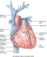
Coronary Circulation
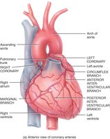
The heart muscle (myocardium) receives its own blood supply through the coronary arteries and is drained by the coronary veins. This circulation is essential for the heart's function.
Coronary Arteries: Branch off the aorta above the aortic semilunar valve.
Left Coronary Artery: Divides into the circumflex branch (supplies left atrium and ventricle) and anterior interventricular artery (supplies both ventricles).
Right Coronary Artery: Divides into the marginal branch (supplies right ventricle) and posterior interventricular artery (supplies both ventricles).
Coronary Veins: Collect waste from cardiac muscle and drain into the coronary sinus, which empties into the right atrium.
Example: Blockage of the coronary arteries can lead to myocardial infarction (heart attack).
Blood Vessels: Structure and Function
Arteries and Veins: Wall Structure
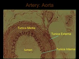
Arteries and veins have similar basic wall structures but differ in thickness and composition due to their functions. Both have three main layers: tunica intima, tunica media, and tunica externa (adventitia).
Artery Wall Layers:
Tunica Intima: Endothelium, subendothelial connective tissue, internal elastic lamina.
Tunica Media: Mainly circular smooth muscle cells and connective tissue fibers.
Tunica Externa: Lamina elastica externa, elastic fibrous layer, connective tissue, vasa vasorum.
Vein Wall Layers:
Tunica Intima: Endothelium, subendothelial connective tissue, internal elastic lamina.
Tunica Media: Smooth muscle cells and loose connective tissue fibers.
Tunica Externa: Loose connective tissue, smooth muscle cells, vasa vasorum.
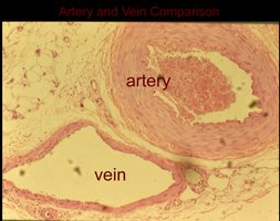
Example: Arteries have thicker tunica media to withstand higher pressure; veins have thinner walls and valves to prevent backflow.
Major Arteries and Veins of the Body
The body contains numerous arteries and veins, each with specific roles in blood transport. Major arteries include the aorta, carotid, subclavian, and iliac arteries. Major veins include the superior and inferior vena cava, brachiocephalic, jugular, and portal veins.
Arteries: Carry blood away from the heart under high pressure.
Veins: Return blood to the heart under lower pressure; often contain valves.
Example: The hepatic portal vein carries nutrient-rich blood from the digestive organs to the liver.
Venous Valves and Varicose Veins
Veins, especially in the limbs, contain valves formed by duplications of the tunica intima. These valves prevent backflow and ensure unidirectional blood flow toward the heart. Dysfunctional valves can lead to varicose veins, where blood pools and the vessel becomes dilated.
Valve Function: Properly functioning valves open and close to prevent backflow.
Varicose Veins: Occur when valves fail, causing vessel dilation and inefficient blood return.
Example: The muscle pump assists venous return by compressing veins during movement.
Summary Table: Comparison of Arteries and Veins
Feature | Arteries | Veins |
|---|---|---|
Wall Thickness | Thick (especially tunica media) | Thin |
Pressure | High | Low |
Valves | Absent | Present (especially in limbs) |
Direction of Blood Flow | Away from heart | Toward heart |
Elasticity | High | Low |
Key Equations
Blood Pressure Equation:
Cardiac Output Equation:
Additional info:
Academic context was added to clarify the structure and function of the cardiovascular system, including the role of valves and the comparison table.
Images were included only when directly relevant to the explanation of the paragraph.
