 Back
BackThe Cardiovascular System: Structure, Function, and Physiology of the Heart
Study Guide - Smart Notes
 Tailored notes based on your materials, expanded with key definitions, examples, and context.
Overview of the Cardiovascular System
Introduction to the Cardiovascular System
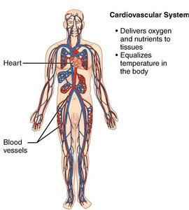
The cardiovascular system is essential for transporting oxygen, nutrients, hormones, and waste products throughout the body. It consists of the heart, blood vessels, and blood, working together to maintain homeostasis and support cellular function.
Heart: Muscular organ that pumps blood.
Blood vessels: Tubular structures (arteries, veins, capillaries) that carry blood throughout the body.
Blood: Fluid that transports gases, nutrients, and waste products.
Main functions: Deliver oxygen and nutrients, remove waste, regulate temperature, and maintain pH balance.
Anatomy of the Heart
Location, Size, and Structure of the Heart
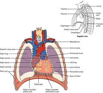
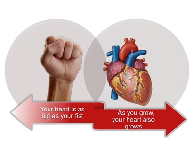
The heart is located slightly to the left of the midline in the thoracic cavity, posterior to the sternum and within the mediastinum. It rests on the diaphragm and is roughly the size of a closed fist, weighing between 250-350 grams in adults.
Apex: The pointed end, directed toward the left hip.
Base: The broad, flat portion facing the posterior rib cage.
Size variations: Athlete's hearts may be larger; pathological enlargement can occur in disease.
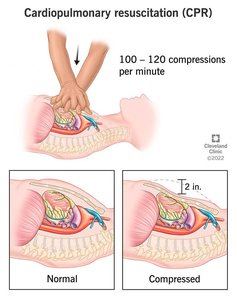
Cardiopulmonary Resuscitation (CPR)
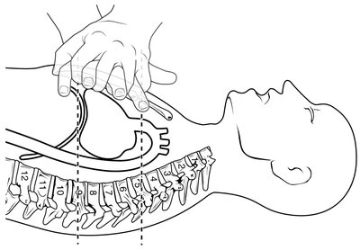
CPR is an emergency technique used to manually pump blood during cardiac arrest, maintaining circulation to vital organs until normal heart function is restored.
Compress the chest at least 2 inches deep at a rate of 100-120 compressions per minute.
Critical for preventing brain damage during cardiac arrest.
Chambers and Valves of the Heart
Heart Chambers
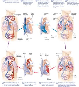
The heart contains four chambers: two atria (superior) and two ventricles (inferior). Blood enters the atria and exits through the ventricles. Valves prevent backflow and ensure unidirectional flow.
Right atrium: Receives deoxygenated blood from the body via the superior and inferior vena cava.
Right ventricle: Pumps blood to the lungs via the pulmonary arteries.
Left atrium: Receives oxygenated blood from the lungs via the pulmonary veins.
Left ventricle: Pumps oxygenated blood to the body via the aorta.
Heart Valves
Valves ensure one-way flow of blood through the heart:
Atrioventricular (AV) valves: Tricuspid (right) and bicuspid/mitral (left) valves separate atria from ventricles.
Semilunar valves: Pulmonary (right) and aortic (left) valves prevent backflow into the ventricles.
Heart Wall and Membranes
Layers of the Heart Wall
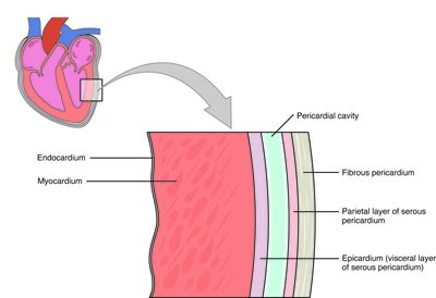
The heart wall consists of three layers:
Epicardium (visceral pericardium): Outer layer, part of the serous membrane.
Myocardium: Middle, muscular layer responsible for contraction.
Endocardium: Inner lining of the heart chambers, composed of simple squamous epithelium (endothelium).
Pericardium and Pericardial Cavity
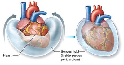
The pericardium is a double-walled sac surrounding the heart, consisting of:
Fibrous pericardium: Tough outer layer that anchors the heart and prevents overfilling.
Serous pericardium: Thin inner layer with parietal and visceral (epicardium) layers, producing serous fluid for lubrication.
Pericardial cavity: Space between serous layers, filled with serous fluid to reduce friction during heartbeats.
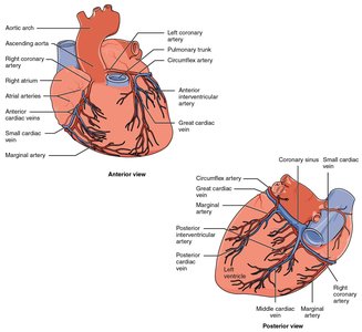
Coronary Circulation
Blood Supply to the Heart
The myocardium is too thick for nutrients to diffuse from the chambers, so the heart has its own blood supply via the coronary arteries and veins.
Coronary arteries: Branch from the aorta to supply oxygenated blood to the heart muscle.
Coronary veins: Collect deoxygenated blood from the myocardium and return it to the right atrium.
Cardiac Muscle and Electrophysiology
Cardiac Muscle Structure and Function
Cardiac muscle cells are striated, branched, and interconnected by intercalated discs, which allow rapid transmission of electrical impulses and coordinated contraction.
Autorhythmicity: The heart generates its own action potentials via pacemaker cells.
Intercalated discs: Specialized connections that facilitate electrical and mechanical coupling between cells.
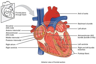
Cardiac Conduction System
The heart's electrical system ensures coordinated contraction:
Sinoatrial (SA) node: Pacemaker, initiates heartbeat.
Atrioventricular (AV) node: Delays impulse, allowing atria to contract before ventricles.
AV bundle (Bundle of His), bundle branches, Purkinje fibers: Distribute impulse through ventricles.
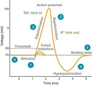
Electrophysiological Terminology
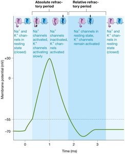
Resting membrane potential: The electrical charge difference across the cell membrane at rest.
Action potential: Rapid change in membrane potential that triggers muscle contraction.
Depolarization: Membrane potential becomes more positive.
Repolarization: Membrane potential returns to negative.
Refractory period: Time during which the cell cannot be re-excited.
The Electrocardiogram (ECG)
Understanding the ECG
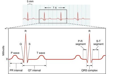
An electrocardiogram (ECG or EKG) records the electrical activity of the heart. It is used to diagnose arrhythmias, myocardial infarction, and other cardiac conditions.
P wave: Atrial depolarization.
QRS complex: Ventricular depolarization (and atrial repolarization).
T wave: Ventricular repolarization.
Mechanical Physiology of the Heart: The Cardiac Cycle
The Cardiac Cycle
The cardiac cycle is the sequence of events in one heartbeat, including contraction (systole) and relaxation (diastole) of the atria and ventricles.
Systole: Contraction phase, blood is pumped out of the chambers.
Diastole: Relaxation phase, chambers fill with blood.
Cardiac Cycle Events
Atrial systole: Atria contract, pushing blood into ventricles.
Ventricular systole: Ventricles contract, ejecting blood into the pulmonary artery and aorta.
Diastole: Chambers relax and fill with blood.
Cardiac Output and Regulation
Cardiac Output
Cardiac output (CO) is the volume of blood pumped by each ventricle per minute. It is a key indicator of heart function and tissue perfusion.
Formula:
Normal CO is about 5 L/min in a healthy adult at rest.
Stroke volume (SV): Volume of blood pumped per beat (typically 70 mL).
Heart rate (HR): Beats per minute (typically 60-100 bpm in adults).
Factors Affecting Cardiac Output
Preload (EDV): Volume of blood in ventricles at end of diastole; increased preload increases SV.
Afterload: Resistance the ventricles must overcome to eject blood; increased afterload decreases SV.
Contractility: Strength of ventricular contraction; increased contractility increases SV.
Autonomic nervous system: Sympathetic stimulation increases HR and contractility; parasympathetic decreases HR.
Equations
Stroke Volume:
Cardiac Output:
Summary Table: Key Features of the Heart
Feature | Description |
|---|---|
Chambers | 2 atria (receive blood), 2 ventricles (pump blood) |
Valves | Tricuspid, bicuspid (mitral), pulmonary, aortic |
Wall Layers | Epicardium, myocardium, endocardium |
Conduction System | SA node, AV node, bundle of His, bundle branches, Purkinje fibers |
Cardiac Output | CO = HR × SV |
Additional info: This guide covers the structure and function of the heart, the cardiac cycle, and the basics of cardiac electrophysiology, as outlined in a typical college-level Anatomy and Physiology course (Chapter 17: The Cardiovascular System: The Heart).
