Back
BackThe Cardiovascular System: Structure, Function, and Physiology
Study Guide - Smart Notes
 Tailored notes based on your materials, expanded with key definitions, examples, and context.
The Cardiovascular System
General Function
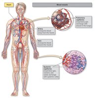
The cardiovascular system consists of the heart and blood vessels, and its primary function is to transport blood throughout the body. This system delivers oxygen and nutrients to tissues while removing carbon dioxide and metabolic wastes. Adequate perfusion, defined as the delivery of blood per time per gram of tissue (mL/min/g), is essential for maintaining cellular health. Continuous heart pumping and healthy, open vessels are required for proper perfusion.
Perfusion: The process of blood delivery to tissues, crucial for cell survival.
Key roles: Oxygen and nutrient delivery, waste removal, and maintenance of tissue health.
Overview of Components
The cardiovascular system is composed of three main types of blood vessels: arteries, veins, and capillaries. Arteries carry blood away from the heart, veins return blood to the heart, and capillaries are the sites of exchange between blood and tissues or air in the lungs.
Arteries: Most carry oxygenated blood, except pulmonary arteries.
Veins: Most carry deoxygenated blood, except pulmonary veins.
Capillaries: Sites of gas, nutrient, and waste exchange.
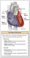
Heart Structure: Chambers and Sides
The heart has two sides, each with an atrium and a ventricle. The right side receives deoxygenated blood and pumps it to the lungs, while the left side receives oxygenated blood and pumps it to the body. The atria are superior chambers that receive blood, and the ventricles are inferior chambers that pump blood away.
Right side: Receives and pumps deoxygenated blood to lungs.
Left side: Receives and pumps oxygenated blood to systemic tissues.
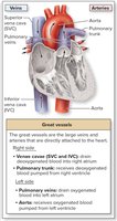
Great Vessels
Great vessels transport blood to and from the heart's chambers. The superior and inferior vena cava drain deoxygenated blood into the right atrium. The pulmonary trunk carries blood from the right ventricle to the lungs, while pulmonary veins return oxygenated blood to the left atrium. The aorta carries blood from the left ventricle to the body.
Superior/inferior vena cava: Veins draining into right atrium.
Pulmonary trunk: Artery carrying deoxygenated blood to lungs.
Pulmonary veins: Veins returning oxygenated blood to left atrium.
Aorta: Artery carrying oxygenated blood to systemic circulation.
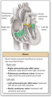
Heart Valves
Two sets of heart valves ensure one-way blood flow: atrioventricular (AV) valves and semilunar valves. AV valves are located between atria and ventricles, while semilunar valves are at the boundary of ventricles and arterial trunks.
Right AV valve (tricuspid): Between right atrium and ventricle.
Left AV valve (bicuspid/mitral): Between left atrium and ventricle.
Pulmonary semilunar valve: Between right ventricle and pulmonary trunk.
Aortic semilunar valve: Between left ventricle and aorta.
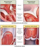
Mechanism of Heart Valves
Heart valves prevent backflow and ensure unidirectional blood flow. AV valves close when ventricles contract, and papillary muscles with tendinous cords prevent inversion. Semilunar valves open when ventricles contract and close when ventricles relax, responding to pressure changes.
AV valves: Prevent backflow to atria; supported by papillary muscles and tendinous cords.
Semilunar valves: Prevent backflow to ventricles; open and close based on pressure gradients.
Pulmonary and Systemic Circulation
The heart supports two circulations: pulmonary and systemic. Pulmonary circulation moves deoxygenated blood from the right heart to the lungs for gas exchange, then returns oxygenated blood to the left heart. Systemic circulation delivers oxygenated blood from the left heart to body tissues and returns deoxygenated blood to the right heart.
Pulmonary circulation: Right heart → lungs → left heart.
Systemic circulation: Left heart → body tissues → right heart.
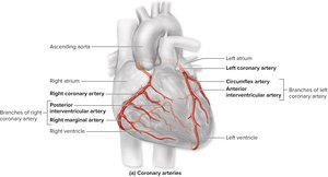
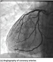
Coronary Circulation
Coronary Arteries and Veins
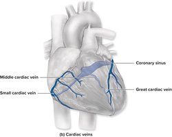
Coronary circulation supplies the heart wall with blood. Coronary arteries deliver oxygenated blood, while coronary veins remove deoxygenated blood. Blockage of coronary arteries can lead to insufficient blood flow and myocardial infarction.
Coronary arteries: Right and left coronary arteries, circumflex, anterior and posterior interventricular arteries.
Coronary veins: Great cardiac vein, middle cardiac vein, small cardiac vein, coronary sinus.
Microscopic Structure and Metabolism of Cardiac Muscle
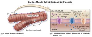
Cardiac Muscle Cells
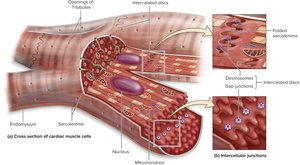
The myocardium is composed of short, branched cardiac muscle cells with one or two central nuclei. These cells are supported by endomysium and are connected by intercalated discs, which include desmosomes (mechanical connection) and gap junctions (electrical connection).
Intercalated discs: Allow coordinated contraction (functional syncytium).
Striated appearance: Due to sarcomere organization.
Metabolism
Cardiac muscle has a high demand for energy, relying primarily on aerobic metabolism. It contains numerous mitochondria, myoglobin, and creatine kinase, and can use various fuel molecules (fatty acids, glucose, lactic acid, amino acids, ketone bodies).
Aerobic metabolism: Makes cardiac muscle susceptible to ischemia.
Heart Conduction System and Innervation
Conduction System
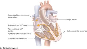
The heart's conduction system initiates and propagates electrical impulses to ensure proper timing of contractions. The SA node (pacemaker) initiates the heartbeat, followed by the AV node, AV bundle, bundle branches, and Purkinje fibers.
SA node: Initiates heartbeat.
AV node: Delays impulse, allowing ventricular filling.
AV bundle and branches: Conduct impulse to ventricles.
Purkinje fibers: Distribute impulse through ventricles.
Innervation of the Heart
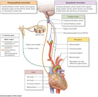
The heart is innervated by both sympathetic and parasympathetic pathways. The cardiac center in the medulla oblongata receives input from baroreceptors and chemoreceptors and sends signals to modify heart rate and force of contraction.
Parasympathetic: Decreases heart rate via vagus nerve.
Sympathetic: Increases heart rate and force via spinal nerves.
Electrical Activity and Cardiac Muscle Contraction
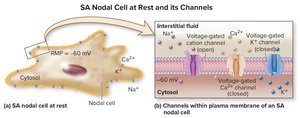
SA Nodal Cells
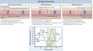
SA nodal cells exhibit autorhythmicity, spontaneously depolarizing to generate action potentials. The resting membrane potential is about −60 mV, and these cells have unique voltage-gated channels that allow them to reach threshold without external stimulation.
Pacemaker potential: Ability to reach threshold spontaneously.
Key channels: Slow and fast voltage-gated channels for Na+, Ca2+, and K+.
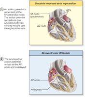
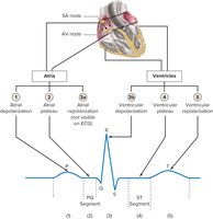
Spread of Action Potential
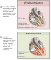
Action potentials initiated at the SA node spread through the atria, reach the AV node (where they are delayed), then pass through the AV bundle, bundle branches, and Purkinje fibers to the ventricles. This ensures coordinated contraction of the heart chambers.
AV node delay: Allows ventricles to fill before contraction.
Ventricular contraction: Begins at apex and moves superiorly.
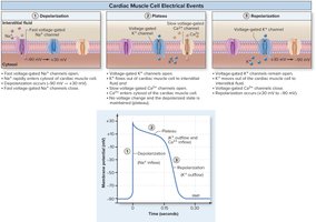
Cardiac Muscle Cell Electrical Events
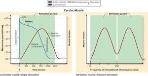
Cardiac muscle cells have a resting membrane potential of −90 mV. Action potentials involve depolarization (Na+ influx), plateau (Ca2+ influx and K+ efflux), and repolarization (K+ efflux). The plateau phase leads to a long refractory period, preventing tetany.
Depolarization: Fast Na+ channels open.
Plateau: Ca2+ enters, K+ leaves.
Repolarization: Ca2+ channels close, K+ channels remain open.
Electrocardiogram (ECG)
ECG Waves and Segments
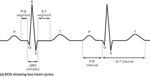
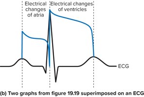
An ECG records the electrical activity of the heart. The P wave reflects atrial depolarization, the QRS complex reflects ventricular depolarization (and atrial repolarization), and the T wave reflects ventricular repolarization. The P-Q and S-T segments correspond to plateau phases.
P wave: Atrial depolarization.
QRS complex: Ventricular depolarization.
T wave: Ventricular repolarization.
P-Q segment: Atrial plateau.
S-T segment: Ventricular plateau.
The Cardiac Cycle
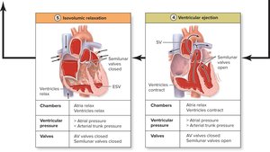
Phases of the Cardiac Cycle
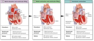
The cardiac cycle includes systole (contraction) and diastole (relaxation). Blood moves down its pressure gradient, and valves ensure forward flow. Key events include atrial relaxation and ventricular filling, atrial contraction, isovolumic contraction, ventricular ejection, and isovolumic relaxation.
End-diastolic volume (EDV): Volume in ventricle at end of filling.
Stroke volume (SV): Volume ejected per beat.
End-systolic volume (ESV): Volume remaining after contraction.
Relationship:
Cardiac Output and Its Regulation
Cardiac Output
Cardiac output (CO) is the amount of blood pumped by a single ventricle in one minute. It is a measure of cardiovascular effectiveness and is determined by heart rate (HR) and stroke volume (SV).
Formula:
Example:
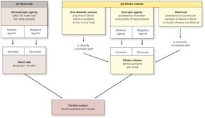
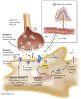
Variables Influencing Heart Rate
Chronotropic agents alter heart rate by affecting nodal cells. Positive agents (e.g., sympathetic stimulation, epinephrine, thyroid hormone, nicotine, cocaine, caffeine) increase heart rate. Negative agents (e.g., parasympathetic stimulation, beta-blockers) decrease heart rate.
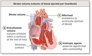
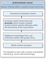
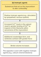
Variables Influencing Stroke Volume
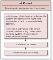
Stroke volume is influenced by end-diastolic volume (EDV), inotropic agents, and afterload. Venous return determines EDV and preload. The Frank-Starling law states that increased EDV leads to greater force of contraction. Inotropic agents alter contractility, and afterload is the resistance to ejection.
Factors Affecting Cardiac Output
Cardiac output varies directly with heart rate and stroke volume. Heart rate is influenced by chronotropic agents, while stroke volume depends on the state of the myocardium, venous return, inotropic agents, and afterload.