 Back
BackThe Cardiovascular System: The Heart – Structure, Function, and Physiology
Study Guide - Smart Notes
 Tailored notes based on your materials, expanded with key definitions, examples, and context.
The Cardiovascular System: The Heart
Introduction to the Cardiovascular System
The cardiovascular system is essential for transporting nutrients, gases, hormones, and wastes throughout the body. It consists of the heart, blood vessels, and blood. The heart acts as a muscular pump, propelling blood through two main circuits: the pulmonary and systemic circuits.
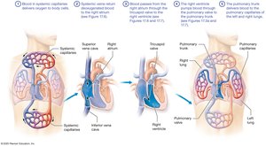
Pulmonary Circuit: Carries deoxygenated blood from the right side of the heart to the lungs for gas exchange and returns oxygenated blood to the left side of the heart.
Systemic Circuit: Delivers oxygenated blood from the left side of the heart to the body and returns deoxygenated blood to the right side of the heart.
Blood Vessel Types
Arteries: Carry blood away from the heart.
Veins: Carry blood toward the heart.
Capillaries: Tiny vessels where exchange of materials occurs between blood and interstitial fluid.
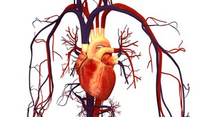
Location and Basic Structure of the Heart
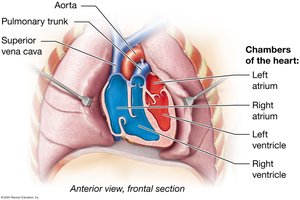
Heart Chambers and Circuits
The heart is divided into four chambers:
Right Atrium: Receives deoxygenated blood from the systemic circuit.
Right Ventricle: Pumps blood into the pulmonary circuit.
Left Atrium: Receives oxygenated blood from the pulmonary circuit.
Left Ventricle: Pumps blood into the systemic circuit.
Heart Location and Anatomy
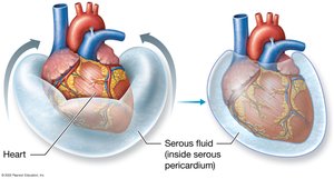
The heart is about the size of a fist, located in the mediastinum within the pericardial cavity. The superior edge is called the base, and the pointed tip is the apex. The heart is surrounded by the pericardium, which consists of:
Parietal Pericardium: Outer layer lining the pericardial sac.
Visceral Pericardium (Epicardium): Inner layer covering the heart surface.
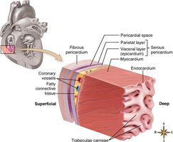
The Heart Wall and Skeleton
Layers of the Heart Wall
Epicardium: Outer layer (visceral pericardium).
Myocardium: Thick, muscular middle layer composed of cardiac muscle cells (myocytes).
Endocardium: Inner layer made of endothelial cells, forming a barrier and regulating electrolyte concentration.
The fibrous skeleton of the heart provides structural support, especially around the valves and septa.
The Great Vessels, Chambers, and Valves of the Heart
Major Vessels
Superior and Inferior Vena Cava: Return deoxygenated blood to the right atrium.
Pulmonary Trunk and Arteries: Carry deoxygenated blood from the right ventricle to the lungs.
Pulmonary Veins: Return oxygenated blood from the lungs to the left atrium.
Aorta: Distributes oxygenated blood from the left ventricle to the body.
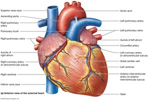
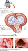
Heart Valves
Atrioventricular (AV) Valves: Between atria and ventricles; prevent backflow during ventricular contraction.
Right AV Valve (Tricuspid): Three cusps between right atrium and ventricle.
Left AV Valve (Bicuspid/Mitral): Two cusps between left atrium and ventricle.
Semilunar (SL) Valves: At the bases of the large arteries leaving the ventricles; prevent backflow into the heart.
Pulmonary Valve: At the base of the pulmonary trunk.
Aortic Valve: At the base of the aorta.
Blood Flow Through the Heart
Pathway of Blood
Deoxygenated blood enters the right atrium via the superior and inferior vena cava.
Passes through the tricuspid valve into the right ventricle.
Pumped through the pulmonary valve into the pulmonary trunk and arteries to the lungs.
Oxygenated blood returns via pulmonary veins to the left atrium.
Passes through the bicuspid (mitral) valve into the left ventricle.
Pumped through the aortic valve into the aorta and systemic circulation.
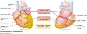
The Coronary Circulation
Blood Supply to the Heart
The heart muscle (myocardium) receives its own blood supply via the coronary arteries and is drained by cardiac veins into the coronary sinus, which empties into the right atrium.
Right Coronary Artery: Supplies right atrium, right ventricle, and parts of the conduction system.
Left Coronary Artery: Supplies left atrium, left ventricle, and interventricular septum.
Coronary Artery Disease and Myocardial Infarction
Coronary Artery Disease (CAD): Buildup of fatty plaques in coronary arteries, reducing oxygen supply to the myocardium.
Myocardial Infarction (Heart Attack): Death of heart muscle due to prolonged lack of oxygen, often from a blocked coronary artery.
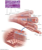
Histology of Cardiac Muscle Tissue
Cardiac Muscle Cell Structure
Small, striated, branched cells with a single central nucleus.
Connected by intercalated discs containing desmosomes (mechanical connection) and gap junctions (electrical connection).
Allows the heart to function as a functional syncytium (coordinated unit).
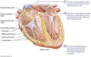
Cardiac Electrophysiology: Pacemaker Cells and the Conduction System
Pacemaker and Conducting Cells
Sinoatrial (SA) Node: Primary pacemaker, initiates heartbeat, located in the right atrium.
Atrioventricular (AV) Node: Delays impulse, allowing atria to contract before ventricles.
AV Bundle, Bundle Branches, Purkinje Fibers: Distribute impulse through ventricles, ensuring coordinated contraction from apex upward.
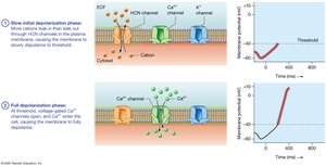
Action Potentials in Pacemaker Cells
Pacemaker cells have an unstable resting potential that gradually depolarizes to threshold, triggering an action potential.
SA node fires at 80–100 times per minute; AV node at 40–60 times per minute.
Cardiac Cycle
Phases of the Cardiac Cycle
Systole: Contraction phase; blood is ejected from a chamber.
Diastole: Relaxation phase; chamber fills with blood.
Blood flows from areas of higher pressure to lower pressure, controlled by the timing of contractions and one-way valves.
Events of the Cardiac Cycle
Atrial Systole: Atria contract, topping off ventricular volume (end-diastolic volume, EDV).
Ventricular Systole: Ventricles contract, AV valves close (isovolumetric contraction), then semilunar valves open for ejection (stroke volume, SV).
Ventricular Diastole: Ventricles relax, semilunar valves close (end-systolic volume, ESV), AV valves open, and passive filling occurs.
Heart Sounds
S1 ("lubb"): Closing of AV valves during ventricular systole.
S2 ("dubb"): Closing of semilunar valves during ventricular diastole.
Heart Murmur: Abnormal sound due to valve regurgitation.
Cardiac Output and Its Regulation
Definitions and Equations
End-Diastolic Volume (EDV): Volume of blood in each ventricle at the end of diastole.
End-Systolic Volume (ESV): Volume of blood remaining in each ventricle after systole.
Stroke Volume (SV): Amount of blood ejected per beat.
Cardiac Output (CO): Volume of blood pumped by each ventricle per minute. where is heart rate (beats/min) and is stroke volume (mL/beat).
Factors Affecting Cardiac Output
Heart Rate (HR): Influenced by autonomic nervous system, hormones, and temperature.
Stroke Volume (SV): Influenced by preload (degree of stretch), contractility (force of contraction), and afterload (resistance to ejection).
Factor | Effect on Cardiac Output |
|---|---|
Increased Preload | Increases CO |
Increased Contractility | Increases CO |
Increased Afterload | Decreases CO |
Autonomic Regulation
Sympathetic Stimulation: Increases heart rate and contractility (norepinephrine release).
Parasympathetic Stimulation: Decreases heart rate (acetylcholine release).
Hormonal Regulation
Thyroid Hormone, Glucagon: Increase heart rate and contractility.
Aldosterone, Antidiuretic Hormone: Increase blood volume and preload.
Atrial Natriuretic Peptide (ANP): Decreases blood volume and preload.
Summary Table: Key Structures and Functions of the Heart
Structure | Function |
|---|---|
Right Atrium | Receives deoxygenated blood from body |
Right Ventricle | Pumps blood to lungs |
Left Atrium | Receives oxygenated blood from lungs |
Left Ventricle | Pumps blood to body |
AV Valves | Prevent backflow into atria |
Semilunar Valves | Prevent backflow into ventricles |
SA Node | Pacemaker of the heart |
Coronary Arteries | Supply blood to myocardium |
Additional info: This guide covers the structure, function, and physiology of the heart as outlined in a typical college-level Anatomy and Physiology course, focusing on Chapter 17: The Cardiovascular System – The Heart.
