 Back
BackThe Cardiovascular System: The Heart – Study Notes
Study Guide - Smart Notes
 Tailored notes based on your materials, expanded with key definitions, examples, and context.
The Cardiovascular System: The Heart
Anatomy of the Heart
The heart is a muscular organ located in the thoracic cavity, responsible for pumping blood throughout the body. It is positioned in the mediastinum, slightly left of the midline, and is protected by the pericardium and thoracic structures.
Location: Lies in the center of the thoracic cavity, in the inferior portion of the mediastinum.
Size: Roughly the size of a fist.
Orientation: Apex points inferiorly toward the left hip; base is directed toward the right shoulder.
Function: Pumps blood to all body tissues, supplying oxygen and nutrients while removing wastes.
Heart Coverings
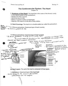
The heart is enclosed in a double-walled sac called the pericardium, which protects and anchors the heart, prevents overfilling, and reduces friction.
Fibrous Pericardium: Outermost layer; tough, dense connective tissue.
Serous Pericardium: Thinner, two-layered membrane (parietal and visceral layers) with pericardial cavity in between, filled with serous fluid to reduce friction.
Heart Wall Layers
The heart wall is composed of three layers, each with distinct structure and function:
Epicardium: Outermost layer; also known as the visceral layer of the serous pericardium.
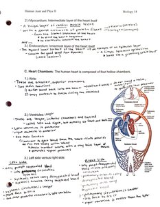
Myocardium: Middle, muscular layer; responsible for contraction and pumping action. Composed of cardiac muscle cells arranged in spiral bundles.
Endocardium: Innermost layer; thin, smooth lining of heart chambers and valves, continuous with blood vessel endothelium.
Heart Chambers
The human heart consists of four hollow chambers: two atria (superior) and two ventricles (inferior). These chambers work together to ensure unidirectional blood flow through the heart and to the body.
Atria: Receive blood returning to the heart; right atrium receives deoxygenated blood from the body, left atrium receives oxygenated blood from the lungs.
Ventricles: Pump blood out of the heart; right ventricle sends blood to the lungs (pulmonary circuit), left ventricle sends blood to the body (systemic circuit).
Blood Flow through the Heart
Circulation of Blood
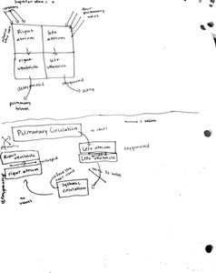
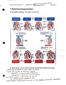
Blood flows through the heart in a specific sequence, ensuring separation of oxygenated and deoxygenated blood. The right side of the heart pumps blood to the lungs (pulmonary circuit), while the left side pumps blood to the rest of the body (systemic circuit).
Deoxygenated blood enters the right atrium via the superior and inferior vena cava.
Blood flows to the right ventricle, then is pumped to the lungs via the pulmonary arteries.
Oxygenated blood returns to the left atrium via the pulmonary veins.
Blood flows to the left ventricle, then is pumped to the body via the aorta.
Heart Valves
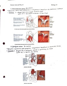
Valves ensure unidirectional blood flow through the heart by opening and closing in response to pressure changes.
Atrioventricular (AV) Valves: Located between atria and ventricles; prevent backflow into atria when ventricles contract. Includes the tricuspid (right) and bicuspid/mitral (left) valves.
Semilunar (SL) Valves: Located at the bases of large arteries leaving the ventricles; prevent backflow into ventricles. Includes the pulmonary (right) and aortic (left) valves.
Valve Disorders
Valve disorders can disrupt normal blood flow and lead to clinical symptoms:
Valve Prolapse: Valve is not fully closed, allowing backflow.
Valve Incompetence: Valve does not close properly, causing regurgitation.
Valve Stenosis: Valve opening is narrowed, restricting blood flow.
Coronary Circulation
The heart muscle (myocardium) receives its own blood supply via the coronary arteries. Blockage of these vessels can lead to myocardial infarction (heart attack).
Right and Left Coronary Arteries: Branch from the aorta and supply oxygenated blood to the heart muscle.
Cardiac Veins: Drain deoxygenated blood from the myocardium into the coronary sinus, which empties into the right atrium.
Heart Disorders
Angina Pectoris: Chest pain due to temporary deficiency of blood to the myocardium.
Myocardial Infarction (MI): Heart attack; prolonged blockage of coronary artery leads to death of heart muscle tissue.
Cardiac Cycle
Phases of the Cardiac Cycle
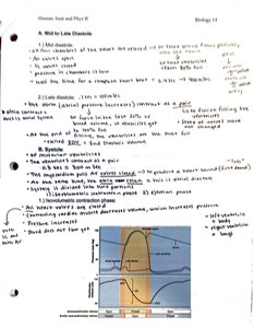
The cardiac cycle consists of all events associated with blood flow through the heart during one heartbeat. It includes alternating periods of contraction (systole) and relaxation (diastole).
Mid to Late Diastole: Ventricles fill with blood as AV valves are open; atria contract at the end of this phase.
Ventricular Systole: Ventricles contract, AV valves close, and blood is ejected into the arteries.
Early Diastole: Ventricles relax, semilunar valves close, and the cycle repeats.
Cardiac Muscle and Electrophysiology
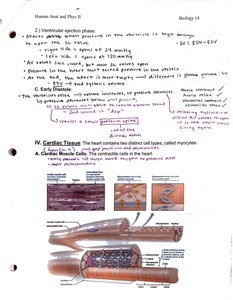
Cardiac Muscle Structure
Cardiac muscle cells (cardiomyocytes) are striated, branched, and interconnected by intercalated discs, which allow for synchronized contraction. These cells contain abundant mitochondria and are adapted for continuous rhythmic activity.
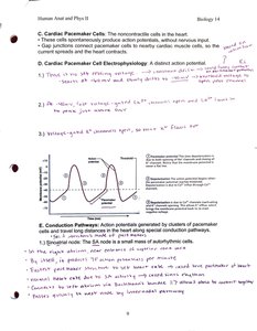
Cardiac Muscle Cell Electrophysiology
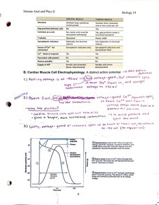
Cardiac muscle cells generate action potentials that trigger contraction. The action potential has distinct phases, including rapid depolarization, plateau, and repolarization.
Depolarization: Rapid influx of Na+ ions.
Plateau: Ca2+ influx maintains depolarization.
Repolarization: K+ efflux restores resting potential.
Cardiac Pacemaker Cells
Pacemaker cells in the sinoatrial (SA) node spontaneously generate action potentials, setting the heart's rhythm. These cells are connected to contractile cells via gap junctions, ensuring coordinated contraction.
Conduction Pathways
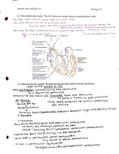
The heart's electrical impulses travel through a specialized conduction system, ensuring efficient and coordinated contraction of the atria and ventricles.
SA Node: Initiates the heartbeat; located in the right atrium.
AV Node: Delays the impulse, allowing atria to contract before ventricles.
AV Bundle (Bundle of His): Conducts impulses from the AV node to the ventricles.
Bundle Branches and Purkinje Fibers: Distribute the impulse throughout the ventricles.
Cardiac Output
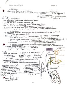
Cardiac output (CO) is the volume of blood pumped by each ventricle per minute. It is calculated as the product of heart rate (HR) and stroke volume (SV):
Formula:
Heart Rate (HR): Number of beats per minute.
Stroke Volume (SV): Volume of blood pumped per beat.
Regulation of Stroke Volume
Stroke volume is influenced by three main factors:
Preload: Degree of stretch of cardiac muscle before contraction (Frank-Starling law).
Contractility: Strength of contraction at a given preload, influenced by sympathetic stimulation and hormones.
Afterload: Pressure the ventricles must overcome to eject blood.
Equation: Where EDV = End-diastolic volume, ESV = End-systolic volume.
Heart Disorders
Tachycardia: Abnormally fast heart rate (>100 bpm).
Bradycardia: Abnormally slow heart rate (<60 bpm).
Congestive Heart Failure (CHF): Heart's pumping efficiency is compromised, leading to fluid accumulation and organ congestion.
Summary Table: Skeletal vs. Cardiac Muscle
The following table compares the properties of skeletal and cardiac muscle:
Feature | Skeletal Muscle | Cardiac Muscle |
|---|---|---|
Location | Attached to bones | Heart wall |
Control | Voluntary | Involuntary |
Cell Shape | Long, cylindrical | Short, branched |
Striations | Present | Present |
Intercalated Discs | Absent | Present |
Contraction | Rapid, short duration | Rhythmic, continuous |
Additional info: These notes provide a comprehensive overview of the heart's anatomy, physiology, and function, suitable for exam preparation in an undergraduate anatomy and physiology course.
