 Back
BackThe Cell: Structure, Function, and Membrane Transport (Chapter 3 Study Notes)
Study Guide - Smart Notes
 Tailored notes based on your materials, expanded with key definitions, examples, and context.
The Cell: Structure, Function, and Membrane Transport
Introduction to Cells
Cells are the basic functional units of life, responsible for maintaining homeostasis in the body. There are two main classes of cells:
Sex Cells (Gametes): Reproductive cells (sperm and oocytes) containing half the number of chromosomes.
Somatic Cells: All other body cells with a full set of chromosomes.
Basic Components of Animal Cells
Plasma Membrane: The outer boundary of the cell, separating the internal environment from the external.
Cytoplasm: The material within the cell, excluding the nucleus. Contains cytosol, organelles, and the cytoskeleton.
Nucleus: The control center of the cell, containing genetic material (DNA).
Plasma Membrane Structure and Function
Phospholipid Bilayer
The plasma membrane is primarily composed of a phospholipid bilayer, which forms a selective barrier between the cell and its environment.
Hydrophilic heads: Face outward toward watery environments.
Hydrophobic tails: Face inward, away from water.
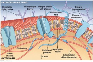
Membrane Proteins and Their Functions
Anchoring Proteins: Stabilize the membrane by attaching to the cytoskeleton or extracellular matrix.
Recognition Proteins: Identify the cell as normal or abnormal.
Enzymes: Catalyze chemical reactions at the membrane surface.
Receptor Proteins: Bind to ligands (e.g., hormones) and trigger cellular responses.
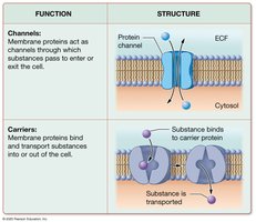
Carrier Proteins: Transport specific substances across the membrane, sometimes requiring ATP.
Channels: Allow passage of water and small solutes.
The Fluid Mosaic Model
The plasma membrane is described by the fluid mosaic model, indicating that its components move laterally within the layer, providing flexibility and dynamic function.
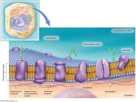
Other Membrane Components
Cholesterol: Stabilizes membrane structure.
Glycolipids and Glycoproteins: Involved in cell recognition and found on the extracellular surface.
Cytoplasm and Organelles
Cytosol
The cytosol is the intracellular fluid containing dissolved proteins, nutrients, ions, and waste products. It differs from extracellular fluid in its high potassium and protein content.
Organelles
Nonmembranous Organelles: Include the cytoskeleton, microvilli, centrioles, cilia, ribosomes, and proteasomes.
Membranous Organelles: Surrounded by membranes; include the endoplasmic reticulum (ER), Golgi apparatus, lysosomes, peroxisomes, and mitochondria.
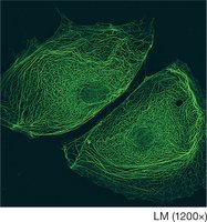
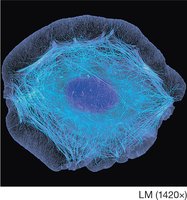
The Cytoskeleton
The cytoskeleton provides structural support, shape, and movement for the cell. It consists of:
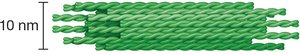
Microfilaments: Thin filaments of actin, providing strength and movement.
Intermediate Filaments: Durable, mid-sized filaments (e.g., keratin) that stabilize organelles and cell position.
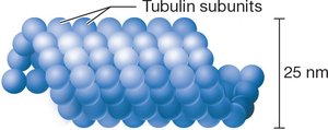
Microtubules: Hollow tubes of tubulin, forming tracks for organelle movement and the spindle apparatus during cell division.
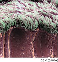
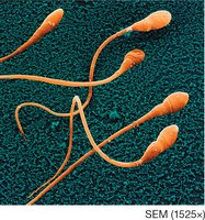
Cilia and Flagella
Cilia are hair-like projections that move fluids across the cell surface, while flagella (e.g., in sperm cells) provide motility.
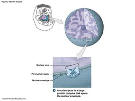
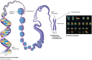
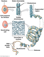
Nucleus
The nucleus is the largest organelle and serves as the cell's control center. It is surrounded by a double membrane (nuclear envelope) with nuclear pores for communication with the cytoplasm.
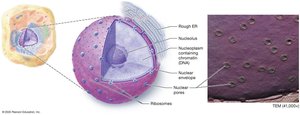
DNA: Contains genetic instructions for building and running the organism.
Chromatin: Loosely coiled DNA in non-dividing cells.
Chromosomes: Tightly coiled DNA in dividing cells.
Nucleolus: Site of ribosomal RNA synthesis and ribosome assembly.
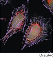
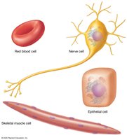
Cell Diversity
Cells vary greatly in size and shape, reflecting their specialized functions in the human body.
Transport Across the Plasma Membrane
Membrane Permeability
The plasma membrane is selectively permeable, allowing some substances to cross while restricting others based on size, charge, shape, and lipid solubility.
Types of Membrane Transport
Passive Transport: Does not require energy; substances move down their concentration gradient.
Active Transport: Requires energy (usually ATP); substances move against their concentration gradient.
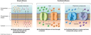
Passive Transport Mechanisms
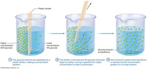
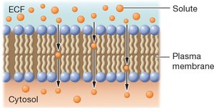
Diffusion: Movement of molecules from high to low concentration due to random motion. Driven by the concentration gradient.
Simple Diffusion: Lipid-soluble molecules and gases pass directly through the membrane.
Channel-Mediated Diffusion: Water and some ions pass through protein channels.
Facilitated Diffusion: Carrier proteins transport large or polar molecules across the membrane.
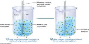
Osmosis
Osmosis is the diffusion of water across a selectively permeable membrane toward a higher solute concentration. Water moves to equalize solute concentrations on both sides of the membrane.
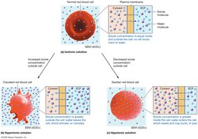
Osmolarity and Tonicity
Isotonic Solution: No net movement of water; cell volume remains constant.
Hypertonic Solution: Higher solute concentration outside the cell; cell loses water and shrinks (crenation).
Hypotonic Solution: Lower solute concentration outside the cell; cell gains water and may burst (lysis).
Active Transport Mechanisms
Primary Active Transport: Uses ATP to move ions against their concentration gradients (e.g., sodium-potassium pump).
Example Equation:
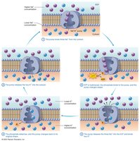
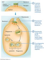
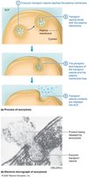
Vesicular Transport: Movement of large particles via vesicles. Includes endocytosis (phagocytosis, pinocytosis, receptor-mediated) and exocytosis.
Summary Table: Plasma Membrane Transport Mechanisms
Mechanism | Process | Factors Affecting Rate | Substances Involved |
|---|---|---|---|
Diffusion | Movement of solutes down concentration gradient | Gradient size, temperature, molecule size | Lipid-soluble molecules, gases |
Osmosis | Movement of water toward higher solute concentration | Osmotic gradient | Water |
Facilitated Diffusion | Carrier proteins transport molecules down gradient | Carrier availability, gradient size | Glucose, amino acids |
Active Transport | Pumps move substances against gradient using ATP | ATP availability, pump number | Ions (Na+, K+, Ca2+) |
Vesicular Transport | Endocytosis/exocytosis via vesicles | ATP availability | Large particles, fluids |
Review and Additional Information
Cells are highly diverse in structure and function, reflecting their specialized roles in the body.
Membrane transport is essential for nutrient uptake, waste removal, and communication with the environment.
Understanding cell structure and membrane transport is foundational for further study in anatomy and physiology.
