 Back
BackThe Cellular Level of Organization: Structure and Function of Cells
Study Guide - Smart Notes
 Tailored notes based on your materials, expanded with key definitions, examples, and context.
Chapter 3: The Cellular Level of Organization
An Introduction to Cells
Cells are the fundamental units of life, forming the basis of all physiological functions in the human body. The cell theory states that all living organisms are composed of cells, all cells arise from pre-existing cells, and cells are the smallest units capable of performing life's essential functions.
Cell Types: Germ cells (sperm and oocyte) are involved in reproduction, while somatic cells make up the rest of the body.
Cellular Homeostasis: Each cell maintains its own homeostasis, contributing to the overall stability of tissues, organs, and systems.
Cell Structure Overview
Cells contain various organelles, each with specialized functions. Organelles are classified as either membranous or nonmembranous, depending on whether they are enclosed by a membrane.
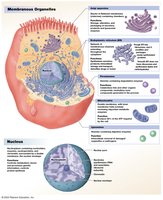
Membranous Organelles: Include the endoplasmic reticulum, Golgi apparatus, lysosomes, peroxisomes, mitochondria, and nucleus.
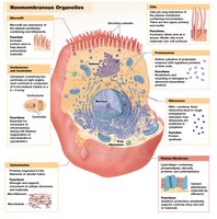
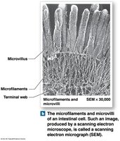
Nonmembranous Organelles: Include the cytoskeleton, centrioles, ribosomes, proteasomes, microvilli, cilia, and flagella.
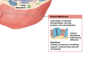
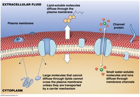
Plasma Membrane
The plasma membrane forms the cell's outer boundary and regulates the movement of substances in and out of the cell. It is composed primarily of lipids and proteins, arranged in a phospholipid bilayer.
Functions: Isolation, protection, sensitivity, support, and regulation of exchange with the environment.
Structure: Hydrophilic heads face outward, hydrophobic tails form the core, cholesterol stabilizes the membrane, and proteins serve various functions.
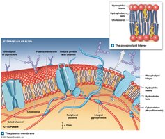
Membrane Proteins and Carbohydrates
Membrane proteins are classified by their location and function. Integral proteins span the membrane, while peripheral proteins are attached to its surface. Membrane carbohydrates form the glycocalyx, which provides protection, cell recognition, and binding specificity.
Types of Proteins: Anchoring, recognition, enzymes, receptor, carrier, and channel proteins.
Glycocalyx: Functions in protection, locomotion, binding, and immune recognition.
Cytoplasm and Nonmembranous Organelles
The cytoplasm contains cytosol (intracellular fluid) and organelles. Nonmembranous organelles provide structural support and facilitate cellular processes.
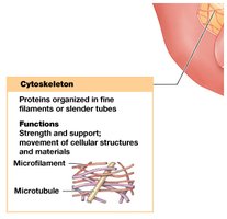
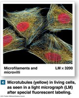
Cytoskeleton: Framework of proteins for shape, strength, and movement.
Microfilaments: Smallest filaments, composed of actin, provide mechanical strength and interact with myosin for muscle contraction.
Intermediate Filaments: Provide strength and stabilize organelle position.
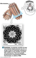
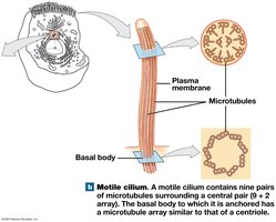
Microtubules: Large, hollow tubes of tubulin, anchor organelles, move vesicles, and form spindle apparatus during cell division.
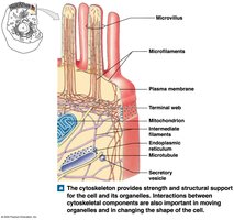
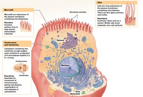
Microvilli, Centrioles, Cilia, and Flagella
These structures extend from the cell surface and play roles in absorption, movement, and cell division.
Microvilli: Increase surface area for absorption.
Centrioles: Organize microtubules and participate in cell division.
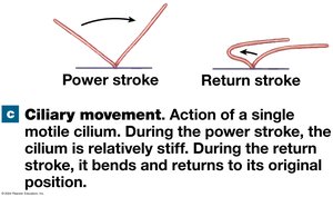
Cilia: Move fluids across cell surfaces; motile cilia have a 9+2 microtubule arrangement.
Flagella: Enable sperm cell movement.
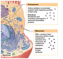
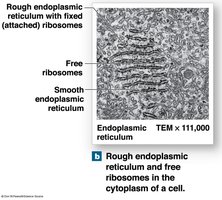
Ribosomes and Proteasomes
Ribosomes synthesize proteins, while proteasomes break down damaged or abnormal proteins.
Ribosomes: Composed of rRNA and proteins; can be free in cytoplasm or attached to ER.
Proteasomes: Hollow cylinders containing proteolytic enzymes for protein degradation.
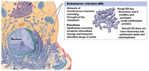
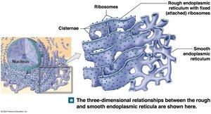
Membranous Organelles
Membranous organelles are enclosed by membranes and perform specialized functions.
Endoplasmic Reticulum (ER): Network of membranes; rough ER synthesizes proteins, smooth ER synthesizes lipids and detoxifies chemicals.
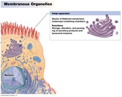
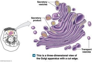
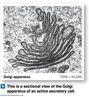
Golgi Apparatus: Modifies, packages, and sorts proteins and lipids for secretion or use within the cell.
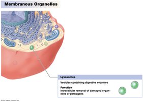
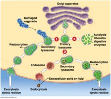
Lysosomes: Contain digestive enzymes for intracellular removal of damaged organelles or pathogens.
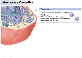
Peroxisomes: Contain enzymes for catabolism of fats and neutralization of toxic compounds.
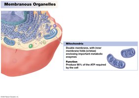
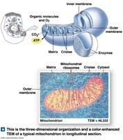
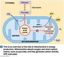
Mitochondria: Produce ATP through cellular respiration; contain their own DNA and ribosomes.
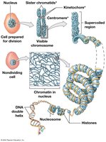
The Nucleus
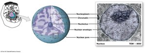
The nucleus is the largest organelle and serves as the control center for cellular operations. It stores genetic information and regulates gene expression.
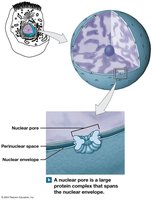
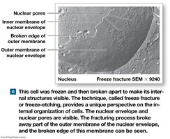
Nuclear Envelope: Double membrane surrounding the nucleus, with nuclear pores for communication.
Nucleoplasm: Fluid inside the nucleus, containing chromatin and nucleoli.
Chromatin and Chromosomes: DNA is loosely coiled as chromatin in non-dividing cells and tightly coiled as chromosomes during cell division.
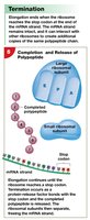
Protein Synthesis
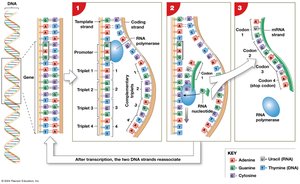
Protein synthesis is the process by which cells build functional polypeptides based on instructions from DNA. It involves transcription and translation.
Transcription: Synthesis of RNA from a DNA template; mRNA carries genetic information from the nucleus to the cytoplasm.
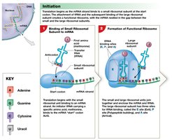
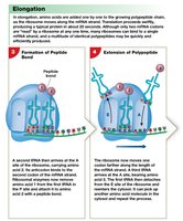
Translation: Ribosomes read mRNA and assemble amino acids into polypeptides; tRNA delivers amino acids to the ribosome.
Genetic Code: Triplet code of DNA bases specifies amino acids; mutations can alter protein structure and function.
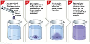
Diffusion and Osmosis
Diffusion and osmosis are passive transport processes that move substances across the plasma membrane.
Diffusion: Net movement of molecules from high to low concentration.
Osmosis: Net diffusion of water across a selectively permeable membrane toward higher solute concentration.
Tonicity: Describes the effect of solute concentration on cell volume (isotonic, hypotonic, hypertonic solutions).
Carrier-Mediated and Vesicular Transport
Cells use specialized mechanisms to transport substances across membranes.
Carrier-Mediated Transport: Uses membrane proteins to move substances; includes facilitated diffusion and active transport.
Vesicular Transport: Moves materials in and out of cells via vesicles; includes endocytosis, exocytosis, and transcytosis.
Membrane Potential
The membrane potential is the electrical potential difference across the plasma membrane, resulting from the unequal distribution of ions. It is essential for nerve impulse transmission and muscle contraction.
Resting Membrane Potential: Typically ranges from −10 mV to −100 mV, with the inside of the cell being more negative.
The Cell Life Cycle
The cell life cycle includes interphase, mitosis, and cytokinesis. These stages are crucial for growth, repair, and maintenance of tissues.
Interphase: Cell grows, duplicates organelles, and replicates DNA.
Mitosis: Division of the nucleus into two identical sets of chromosomes.
Cytokinesis: Division of the cytoplasm, producing two daughter cells.
Regulation of the Cell Life Cycle
Cell division is regulated by internal and external factors. Disruption of regulation can lead to uncontrolled cell growth and cancer.
Regulatory Factors: Internal (MPF), external (growth factors), repressor genes, and telomere length.
Cell Division and Cancer
Cancer results from abnormal cell division due to mutations in genes regulating growth and differentiation. Tumors can be benign or malignant, with malignant tumors capable of metastasis.
Oncogenes: Mutated genes that promote cancer.
Carcinogens: Agents that cause mutations leading to cancer.
Cellular Differentiation
Cellular differentiation is the process by which cells become specialized by turning off certain genes. This allows for the formation of diverse cell types with unique functions.
Importance: Enables the development of specialized tissues and organs.
