 Back
BackThe Cellular Level of Organization: Structure and Function
Study Guide - Smart Notes
 Tailored notes based on your materials, expanded with key definitions, examples, and context.
The Cellular Level of Organization
An Introduction to Cells
Cells are the fundamental units of life, forming the basis of all organisms. The cell theory states that all cells arise from preexisting cells, are the smallest units capable of vital physiological functions, and maintain homeostasis at the cellular level. Cytology, a branch of cell biology, studies cell structure and function. Cells are classified as sex cells (sperm and oocytes) and somatic cells (all other body cells).
Sex cells: Specialized for reproduction.
Somatic cells: Perform all other bodily functions.
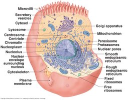
Cell Structure: Model Cell Anatomy
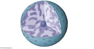
The cell contains various organelles, each with specific functions. The nucleus is the control center, surrounded by a double membrane called the nuclear envelope, containing nuclear pores for communication. The cytoplasm includes cytosol and organelles such as mitochondria, endoplasmic reticulum, Golgi apparatus, lysosomes, and ribosomes.
Nucleus: Stores genetic information and controls metabolism.
Cytoplasm: Contains dissolved nutrients, ions, proteins, and waste products.
Organelles: Structures with specialized functions.
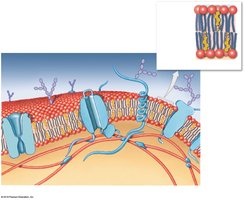
Plasma Membrane
Structure and Function
The plasma membrane separates the cytoplasm from the extracellular fluid, providing physical isolation, regulating exchange, sensitivity to the environment, and structural support. It is composed of a phospholipid bilayer with hydrophilic heads facing outward and hydrophobic tails inward, forming a barrier to ions and water-soluble compounds.
Membrane lipids: Phospholipid bilayer, cholesterol for stability.
Membrane proteins: Anchoring, recognition, enzymes, receptors, carriers, channels.
Membrane carbohydrates: Glycocalyx for lubrication, protection, and recognition.
Organelles within the Cytoplasm
Cytoskeleton
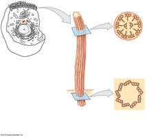
The cytoskeleton provides structural support and shape to the cell. It consists of microfilaments (actin), intermediate filaments, and microtubules (tubulin). Microfilaments interact with myosin for muscle contraction, intermediate filaments provide durability, and microtubules anchor organelles and facilitate movement.
Microfilaments: Mechanical strength, muscle contraction.
Intermediate filaments: Strengthen and stabilize cell.
Microtubules: Change cell shape, move organelles, form centrioles.
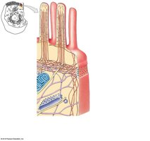
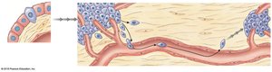
Microvilli, Cilia, and Flagella
Microvilli increase surface area for absorption and attach to the cytoskeleton. Cilia are slender extensions that move fluids across the cell surface; motile cilia are found in respiratory and reproductive tracts, while primary cilia sense environmental stimuli. Flagella are whip-like extensions for cell movement.
Microvilli: Enhance absorption.
Cilia: Move fluids, sense stimuli.
Flagella: Enable cell locomotion.
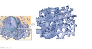
Endoplasmic Reticulum (ER)
The ER is a network of membranes with storage chambers called cisternae. The smooth ER (SER) synthesizes lipids, steroids, and glycogen, while the rough ER (RER) is covered with ribosomes and synthesizes proteins and glycoproteins, folding them into functional structures and packaging them for transport.
SER: Lipid and carbohydrate synthesis.
RER: Protein synthesis and folding.
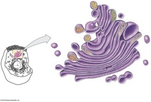
Golgi Apparatus
The Golgi apparatus modifies, packages, and sorts proteins and lipids for secretion or use within the cell. It adds or removes carbohydrates, renews the plasma membrane, and packages enzymes into lysosomes.
Modifies secretions: Hormones, enzymes.
Renews plasma membrane.
Packages enzymes: Lysosomes.
Lysosomes
Lysosomes are enzyme-containing vesicles produced by the Golgi apparatus. Primary lysosomes contain inactive enzymes, while secondary lysosomes form when they fuse with damaged organelles or endosomes, activating enzymes to destroy bacteria, break down molecules, and recycle organelles. Autolysis is the self-destruction of damaged cells.
Primary lysosomes: Inactive enzymes.
Secondary lysosomes: Active enzymes, digestion.
Autolysis: Cell self-destruction.
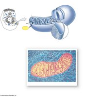
Mitochondria
Mitochondria are the cell's powerhouses, with a smooth outer membrane and folded inner membrane (cristae) surrounding the matrix. They produce ATP through aerobic metabolism, using oxygen to break down glucose. Glycolysis occurs in the cytosol, followed by the citric acid cycle and electron transport chain in the mitochondria.
ATP production: Aerobic metabolism.
Glycolysis: Glucose to pyruvate.
Citric acid cycle: Occurs in matrix.
Electron transport chain: Inner membrane.
Cell Nucleus
Structure and Function
The nucleus is the largest organelle and the cell's control center. It is surrounded by a double membrane (nuclear envelope) with nuclear pores for communication. The nucleus stores genetic information in the form of DNA, organized into genes, which are functional units of heredity.
Nuclear envelope: Double membrane.
Nuclear pores: Communication passages.
Genetic code: DNA instructions, triplet code.
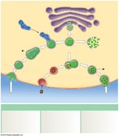
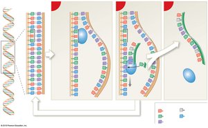
Protein Synthesis
Transcription and Translation
Protein synthesis involves gene activation, DNA replication, transcription (formation of RNA from DNA), and translation (production of proteins from mRNA). Transcription occurs when RNA polymerase binds to DNA, reads the code, and forms mRNA. Translation occurs in the cytoplasm, where ribosomes assemble amino acids into proteins based on mRNA codons.
Transcription: DNA to mRNA.
Translation: mRNA to protein.
Enzymes: Join amino acids with peptide bonds.
Diffusion and Osmosis
Membrane Permeability
The plasma membrane is selectively permeable, allowing some materials to move freely while restricting others based on size, charge, shape, and lipid solubility. Transport can be passive (diffusion, osmosis) or active (carrier-mediated, vesicular).
Passive processes: No energy required.
Active processes: Require energy (ATP).
Diffusion
Diffusion is the net movement of substances from areas of higher concentration to lower concentration, driven by random molecular motion. Factors influencing diffusion include distance, molecule size, temperature, concentration gradient, and electrical forces.
Simple diffusion: Lipid-soluble compounds, gases.
Channel-mediated diffusion: Water-soluble compounds, ions.
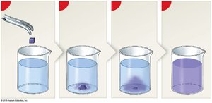
Osmosis
Osmosis is the diffusion of water across a selectively permeable membrane toward a solution with more solutes. Osmotic pressure is the force driving water movement, and hydrostatic pressure blocks osmosis. Osmolarity is the total solute concentration, and tonicity describes how a solution affects cell size and shape.
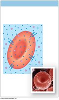
Isotonic solution: No osmotic flow; cell remains unchanged.
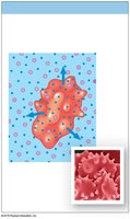
Hypotonic solution: Water enters cell; cell swells and may rupture (hemolysis).
Hypertonic solution: Water leaves cell; cell shrinks (crenation).
Solution Type | Effect on Cell |
|---|---|
Isotonic | No change |
Hypotonic | Swelling, possible rupture |
Hypertonic | Shrinking (crenation) |

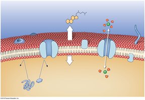
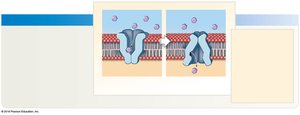
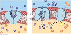
Carrier-Mediated and Vesicular Transport
Carrier-Mediated Transport
Carrier proteins transport ions or organic substrates across the plasma membrane. Transport can be symport (same direction) or antiport (opposite directions). Facilitated diffusion is passive, while active transport requires energy to move substances against concentration gradients.
Facilitated diffusion: Carrier proteins move large molecules (glucose, amino acids).
Active transport: Ion pumps, exchange pumps (e.g., sodium-potassium pump).
Primary active transport: Uses ATP directly.
Secondary active transport: Uses concentration gradients established by ATP.
Vesicular Transport
Vesicular transport moves materials in and out of cells via vesicles. Endocytosis imports materials (receptor-mediated, pinocytosis, phagocytosis), while exocytosis exports materials by fusing vesicles with the plasma membrane.
Endocytosis: Import of extracellular materials.
Exocytosis: Export of cellular products and wastes.
Membrane Potential
Resting Membrane Potential
Membrane potential is the difference in charge across the plasma membrane, typically ranging from –10 mV to –100 mV. It is essential for cell signaling and function.
Cell Life Cycle
Cell Division and Apoptosis
Cell division produces two daughter cells, balancing cell loss. Interphase is the nondividing period, while mitosis and cytokinesis produce new cells. Apoptosis is genetically controlled cell death. The mitotic rate varies by cell type and energy requirements.
Interphase: G0, G1, S, G2 phases; DNA replication.
Mitosis: Prophase, metaphase, anaphase, telophase.
Cytokinesis: Division of cytoplasm.
Regulation of Cell Life Cycle and Cancer
Cell Division Regulation
Cell division is regulated by internal factors (MPF), extracellular growth factors, repressor genes, and telomere length. Faulty repressors can lead to cancer.
Cell Division and Cancer
Cancer results from abnormal cell proliferation due to mutations in genes controlling cell growth. Tumors can be benign (contained) or malignant (invasive). Metastasis is the spread of cancer to other tissues, beginning with invasion and escape into circulation.
Oncogenes: Modified genes causing cancer.
Mutagens: Agents causing mutations.
Carcinogens: Cancer-causing mutagens.
Metastasis: Spread of cancer cells.
Cellular Differentiation
Formation of Specialized Cells
All cells contain the same genetic material, but cellular differentiation occurs by turning off genes not needed by a particular cell type. This process allows for the formation of specialized cells such as liver cells, fat cells, and neurons.
