 Back
BackThe Central Nervous System: Structure and Anatomy of the Brain and Spinal Cord
Study Guide - Smart Notes
 Tailored notes based on your materials, expanded with key definitions, examples, and context.
The Central Nervous System (CNS)
Overview of the CNS
The central nervous system (CNS) consists of the brain and spinal cord. It is responsible for integrating sensory information and coordinating bodily functions. The CNS is protected by bony structures (skull and vertebral column), meninges, and cerebrospinal fluid (CSF).
Brain: The control center for the body, responsible for processing sensory information, generating thoughts, and controlling movements.
Spinal Cord: Conducts signals to and from the brain and controls reflex activities.
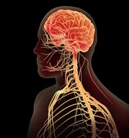
Major Parts of the Brain
Gross Anatomy of the Brain
The brain is divided into several major regions, each with specialized functions. These include the cerebrum, diencephalon, brainstem, and cerebellum.
Cerebrum: Largest part, responsible for higher brain functions such as thought, action, and sensory processing.
Diencephalon: Contains structures such as the thalamus and hypothalamus, involved in sensory relay and homeostasis.
Brainstem: Includes the midbrain, pons, and medulla oblongata; controls vital functions like breathing and heart rate.
Cerebellum: Coordinates voluntary movements and balance.
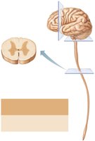
Gray Matter and White Matter in the CNS
Distribution Patterns
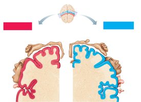
Gray matter consists mainly of neuron cell bodies and is involved in processing and cognition. White matter is composed of myelinated axons that transmit signals between different CNS regions.
Spinal Cord: Central gray matter surrounded by white matter.
Brain: Additional gray matter regions (nuclei) are embedded within white matter, and the cerebrum and cerebellum have an outer gray matter cortex.
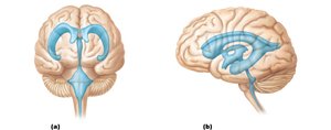
Ventricles of the Brain
Structure and Function
The brain contains interconnected cavities called ventricles, filled with cerebrospinal fluid (CSF). The ventricles cushion the brain, remove waste, and transport nutrients.
Lateral Ventricles: Paired, located in each cerebral hemisphere.
Third Ventricle: Located in the diencephalon.
Fourth Ventricle: Located between the brainstem and cerebellum.
Cerebral Aqueduct: Connects the third and fourth ventricles.
The Cerebrum
Structure and Functional Areas
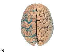
The cerebrum is divided into two hemispheres connected by the corpus callosum. The surface is highly folded, increasing surface area for cortical neurons. The cerebral cortex is responsible for conscious thought, voluntary movement, and sensory perception.
Longitudinal Fissure: Separates the two hemispheres.
Corpus Callosum: Major white matter tract connecting hemispheres.

Lobes of the Cerebral Cortex
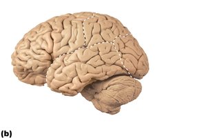
The cerebrum is divided into lobes by sulci (grooves) and fissures. Each lobe has specialized functions:
Frontal Lobe: Voluntary movement, planning, reasoning.
Parietal Lobe: Sensory processing, spatial orientation.
Temporal Lobe: Auditory processing, memory.
Occipital Lobe: Visual processing.
Insular Lobe: Buried under other lobes, involved in taste and visceral sensation.
Functional Maps of the Cerebral Cortex
Motor and Sensory Homunculi
Specific regions of the cerebral cortex correspond to different body parts for motor control and sensory perception. These are represented as 'maps' in the precentral (motor) and postcentral (sensory) gyri.
Primary Motor Cortex: Controls voluntary movements.
Primary Somatosensory Cortex: Receives sensory input from the body.
Internal Structures of the Brain
Midsagittal Anatomy
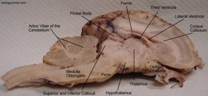
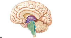
A midsagittal section reveals key internal structures, including the corpus callosum, thalamus, hypothalamus, pineal gland, and ventricles. These structures are essential for communication, hormone regulation, and homeostasis.
Thalamus: Relay station for sensory information.
Hypothalamus: Regulates autonomic and endocrine functions.
Pineal Gland: Secretes melatonin, regulates circadian rhythms.
Fornix: White matter tract involved in memory.
The Diencephalon
Hypothalamic Nuclei
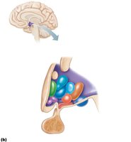
The hypothalamus contains several nuclei that regulate homeostasis, including temperature, hunger, thirst, and circadian rhythms. It also controls the pituitary gland via the infundibulum.
Supraoptic and Paraventricular Nuclei: Produce hormones released by the posterior pituitary.
Suprachiasmatic Nucleus: Controls circadian rhythms.
Mammillary Bodies: Involved in memory processing.
The Brainstem
Cross-Sectional Anatomy
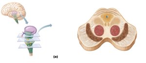
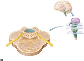
The brainstem consists of the midbrain, pons, and medulla oblongata. It contains important nuclei and tracts for sensory and motor function, as well as centers for vital autonomic functions.
Midbrain: Contains the superior and inferior colliculi (visual and auditory reflexes), substantia nigra, and red nucleus.
Pons: Relays information between the cerebrum and cerebellum; involved in breathing regulation.
Medulla Oblongata: Controls heart rate, blood pressure, and respiration.
The Cerebellum
Structure and Function
The cerebellum coordinates voluntary movements, balance, and posture. It has a highly folded cortex and an internal branching white matter structure called the arbor vitae.
Anterior and Posterior Lobes: Separated by the primary fissure.
Vermis: Central region connecting the two hemispheres.
The Limbic System
Components and Functions
The limbic system is a group of structures involved in emotion, memory, and motivation. It includes parts of the cerebrum and diencephalon.
Amygdala: Processes emotions such as fear and pleasure.
Hippocampus: Essential for memory formation.
Cingulate Gyrus: Involved in emotional regulation and pain processing.
The Reticular Formation
Role in Arousal and Consciousness
The reticular formation is a network of neurons in the brainstem that regulates arousal, alertness, and the sleep-wake cycle. It filters incoming stimuli and helps maintain consciousness.
Reticular Activating System (RAS): Keeps the cerebral cortex alert and conscious.
Brain Waves and Sleep
Electroencephalography (EEG) and Sleep Stages
EEG measures electrical activity in the brain, revealing different types of brain waves associated with various states of consciousness and sleep stages.
Alpha Waves: Awake but relaxed.
Beta Waves: Awake and alert.
Theta Waves: Common in children, also in light sleep.
Delta Waves: Deep sleep.
Meninges and Cerebrospinal Fluid (CSF)
Protective Coverings of the CNS
The CNS is protected by three connective tissue membranes called meninges: dura mater, arachnoid mater, and pia mater. CSF circulates in the subarachnoid space, providing cushioning and nutrient transport.
Dura Mater: Tough, outermost layer.
Arachnoid Mater: Middle, web-like layer.
Pia Mater: Delicate, innermost layer adhering to the CNS surface.
CSF: Produced by the choroid plexus, circulates through ventricles and subarachnoid space, absorbed into venous sinuses via arachnoid granulations.
The Spinal Cord
Gross Structure and Meninges
The spinal cord extends from the foramen magnum to the conus medullaris. It is protected by vertebrae, meninges, and CSF. The spinal cord gives rise to spinal nerves and is organized into cervical, thoracic, lumbar, and sacral regions.
Epidural Space: Between vertebrae and dura mater, contains fat and blood vessels.
Subarachnoid Space: Contains CSF, used for lumbar puncture procedures.
Cauda Equina: Bundle of nerve roots at the lower end of the spinal cord.
Cross-Sectional Anatomy of the Spinal Cord
The spinal cord consists of central gray matter (shaped like a butterfly) surrounded by white matter. The gray matter contains neuron cell bodies, while the white matter contains ascending and descending tracts.
Dorsal Horn: Sensory neurons and interneurons.
Ventral Horn: Motor neurons.
Lateral Horn: Present in thoracic and upper lumbar regions, contains autonomic motor neurons.
Central Canal: Contains CSF.
Organization of Gray Matter
The gray matter of the spinal cord is organized into regions that process sensory and motor information:
Somatic Sensory (SS): Receives input from skin, muscles, and joints.
Visceral Sensory (VS): Receives input from internal organs.
Visceral Motor (VM): Autonomic motor neurons.
Somatic Motor (SM): Motor neurons to skeletal muscle.
White Matter and Spinal Tracts
White matter is organized into columns (funiculi) containing ascending (sensory) and descending (motor) tracts:
Ascending Tracts: Carry sensory information to the brain (e.g., spinothalamic, dorsal columns).
Descending Tracts: Carry motor commands from the brain to the body (e.g., corticospinal, rubrospinal tracts).
Descending Motor Pathways
The brain influences movement through descending pathways, including the pyramidal (corticospinal) tracts, which decussate (cross) in the medulla oblongata and synapse on lower motor neurons in the spinal cord.
Lateral and Ventral Corticospinal Tracts: Control voluntary movements of the limbs and trunk.
Additional info: The sheep brain is commonly used in anatomy labs to study mammalian brain structure due to its similarity to the human brain in gross anatomy, despite differences in size and complexity.
