 Back
The Digestive System: Structure, Function, and Regulation A&P 202 CH 25
Study Guide - Smart Notes
 Tailored notes based on your materials, expanded with key definitions, examples, and context.
The Digestive System
Overview and General Anatomy
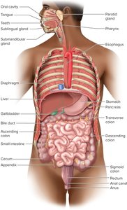
The digestive system is a complex organ system responsible for processing food, extracting nutrients, and eliminating waste. It consists of the digestive tract (alimentary canal) and accessory organs. The tract is a continuous muscular tube extending from the mouth to the anus, while accessory organs assist in digestion through secretion and mechanical processing.
Major Functions: Ingestion, digestion (mechanical and chemical), absorption, compaction, and defecation.
Major Regions: Mouth, pharynx, esophagus, stomach, small intestine, large intestine.
Accessory Organs: Teeth, tongue, salivary glands, liver, gallbladder, pancreas.
Histological Layers of the Digestive Tract
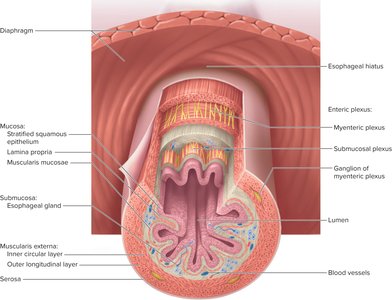
The wall of the digestive tract is organized into four main layers, each with specialized functions:
Mucosa: Innermost layer; consists of epithelium (simple columnar or stratified squamous), lamina propria, and muscularis mucosae. Contains mucosa-associated lymphatic tissue (MALT).
Submucosa: Thicker layer of loose connective tissue containing blood vessels, lymphatics, nerves, and sometimes glands.
Muscularis externa: Usually two layers (inner circular and outer longitudinal); responsible for peristalsis and segmentation. In some regions, forms sphincters.
Serosa (or adventitia): Outermost layer; serosa is a thin areolar tissue with mesothelium, while adventitia is fibrous connective tissue binding organs to surrounding structures.
Enteric Nervous System
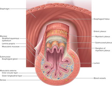
The digestive tract contains its own nervous network, the enteric plexus, which regulates motility, secretion, and blood flow. It can function independently but is influenced by the central nervous system.
Submucosal plexus: Controls glandular secretions and movements of the muscularis mucosae.
Myenteric plexus: Located between muscle layers; controls peristalsis and muscular contractions.
Relationship to the Peritoneum
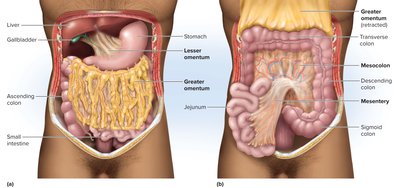
The peritoneum is a serous membrane lining the abdominal cavity and covering abdominal organs. The mesentery is a double layer of peritoneum that supports the intestines and provides a pathway for blood vessels, nerves, and lymphatics.
Intraperitoneal organs: Surrounded by peritoneum on both sides (e.g., stomach, liver).
Retroperitoneal organs: Covered by peritoneum on anterior side only (e.g., duodenum, pancreas).
Omenta: Greater and lesser omentum are extensions of the peritoneum associated with the stomach.
The Oral Cavity and Associated Structures
Anatomy of the Mouth
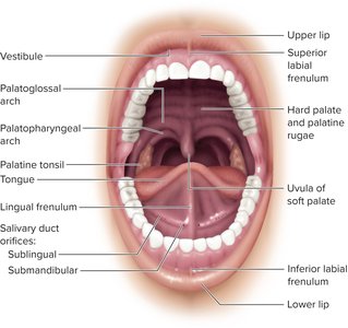
The mouth (oral cavity) is the entry point for food and is involved in ingestion, mechanical digestion, taste, and speech. It is lined by stratified squamous epithelium, keratinized in areas subject to abrasion.
Boundaries: Cheeks, lips, palate, and tongue.
Vestibule: Space between cheeks/lips and teeth.
Palate: Hard (bony) and soft (muscular) palate separate oral and nasal cavities.
The Teeth (Dentition)
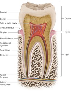
Teeth are essential for mechanical digestion (mastication). Each tooth consists of a crown, neck, and root, and is embedded in an alveolus (tooth socket) by a periodontal ligament.
Regions: Crown (above gum), neck (junction), root (below gum).
Tissues: Enamel (hard, noncellular, covers crown), dentin (bulk of tooth), cement (covers root).
Pulp cavity: Contains nerves and blood vessels.
Types of teeth: Incisors (cutting), canines (tearing), premolars and molars (grinding).
Development of Teeth
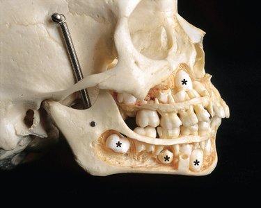
Humans have two sets of teeth: deciduous (baby) teeth and permanent teeth. Deciduous teeth erupt from 6 to 30 months; permanent teeth replace them between ages 6 and 25 years.
Salivary Glands and Saliva
Types and Functions of Salivary Glands
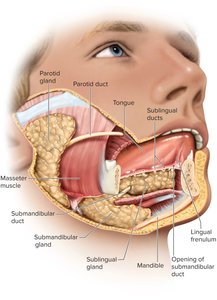
Salivary glands produce saliva, which moistens food, begins starch and fat digestion, cleanses teeth, and inhibits bacterial growth.
Intrinsic glands: Small glands in oral tissues (lingual, labial, palatine, buccal).
Extrinsic glands: Parotid, submandibular, and sublingual glands, each with ducts opening into the oral cavity.
Microscopic Anatomy of Salivary Glands
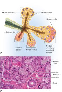
Salivary glands are composed of mucous and serous acini, which secrete different components of saliva. Mixed acini contain both cell types.
Stomach: Structure and Function
Gastric Secretions and Their Regulation
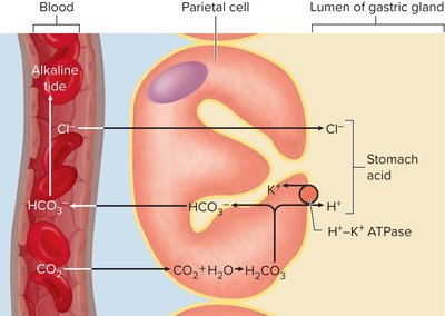
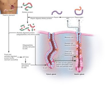
The stomach secretes gastric juice (water, hydrochloric acid, pepsin) and other substances essential for digestion. Parietal cells secrete HCl, which activates enzymes and provides an acidic environment.
Hydrochloric acid (HCl): Produced by parietal cells via the action of carbonic anhydrase and H+/K+ ATPase pumps.
Equation for HCl production:
H+ is pumped into the gastric lumen, Cl- follows, forming HCl.
Pepsin and Protein Digestion
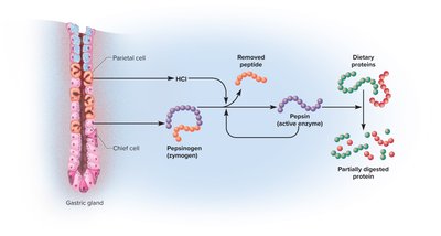
Pepsinogen, secreted by chief cells, is activated to pepsin by HCl. Pepsin digests proteins into shorter peptides, initiating protein digestion in the stomach.
Peptic Ulcers
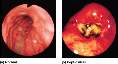
Peptic ulcers are erosions of the stomach wall caused by the action of acid and pepsin, often associated with Helicobacter pylori infection.
Regulation of Gastric Function
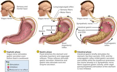
Gastric secretion and motility are regulated by neural and hormonal mechanisms, divided into three overlapping phases:
Cephalic phase: Initiated by sight, smell, taste, or thought of food; mediated by the vagus nerve.
Gastric phase: Triggered by food in the stomach; involves stretch and chemical stimuli, and secretion of acetylcholine, histamine, and gastrin.
Intestinal phase: Begins as chyme enters the duodenum; initially stimulates, then inhibits gastric activity via enterogastric reflex and hormones (secretin, CCK).
Liver, Gallbladder, and Pancreas
Liver: Structure and Function
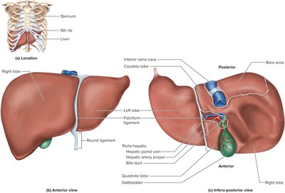
The liver is the largest gland in the body, with four lobes and a complex internal structure. Its main digestive function is the production of bile, which aids in fat digestion.
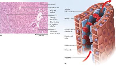
Hepatic lobules: Cylindrical structures containing hepatocytes arranged around a central vein.
Blood supply: Receives nutrient-rich blood from the hepatic portal vein and oxygenated blood from the hepatic artery.
Bile secretion: Bile canaliculi collect bile, which drains into bile ducts and eventually the common hepatic duct.
Gallbladder and Bile
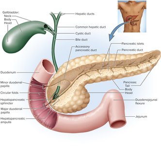
The gallbladder stores and concentrates bile produced by the liver. Bile contains bile acids, cholesterol, and pigments (bilirubin). Bile acids are essential for emulsifying fats in the small intestine.
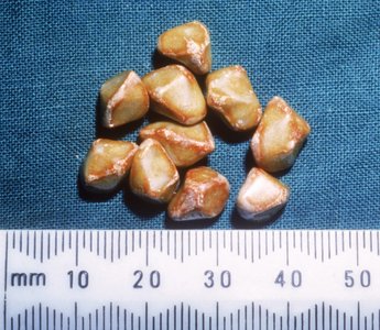
Gallstones: Hard masses formed from cholesterol, calcium carbonate, and bilirubin; can obstruct bile flow and cause jaundice.
Pancreas: Structure and Function
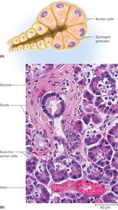
The pancreas is both an endocrine and exocrine gland. Its exocrine portion produces pancreatic juice, which contains digestive enzymes and bicarbonate to neutralize stomach acid in the duodenum.
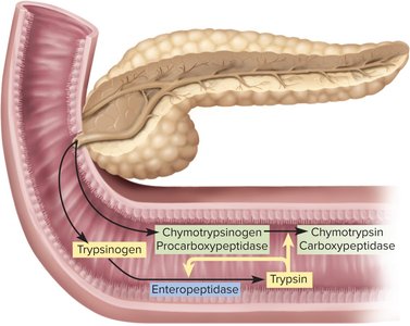
Enzymes: Pancreatic amylase (starch), lipase (fats), proteases (trypsinogen, chymotrypsinogen, procarboxypeptidase).
Activation: Trypsinogen is converted to trypsin by enteropeptidase in the small intestine; trypsin activates other proteases.
Additional info: This summary covers the anatomy, histology, and regulation of the digestive system, focusing on the mouth, teeth, salivary glands, stomach, liver, gallbladder, and pancreas. For further details on the small and large intestines, absorption, and clinical correlations, refer to subsequent sections of the source material.
