 Back
The Digestive System: Structure, Function, and Processes
Study Guide - Smart Notes
 Tailored notes based on your materials, expanded with key definitions, examples, and context.
The Digestive System: Overview
Introduction to the Digestive System
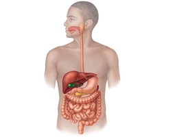
The digestive system is responsible for breaking down food, absorbing nutrients, and eliminating waste. It consists of the alimentary canal (gastrointestinal tract) and accessory digestive organs. The alimentary canal includes the mouth, pharynx, esophagus, stomach, small intestine, and large intestine. Accessory organs include the teeth, tongue, salivary glands, liver, gallbladder, and pancreas.
Major Digestive Processes
Six Essential Activities
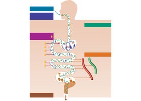
Ingestion: Taking food into the digestive tract.
Propulsion: Moving food through the tract, primarily by swallowing and peristalsis.
Mechanical Digestion: Physical breakdown of food (chewing, churning, segmentation).
Chemical Digestion: Enzymatic breakdown of food molecules into their building blocks.
Absorption: Transport of digested nutrients from the lumen of the GI tract into the blood or lymph.
Defecation: Elimination of indigestible substances as feces.
Motility in the Digestive Tract
Peristalsis and Segmentation
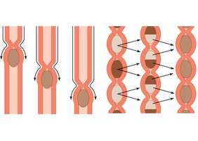
Two primary types of movement occur in the alimentary canal:
Peristalsis: Involuntary, wave-like contractions that propel food forward through the digestive tract.
Segmentation: Rhythmic contractions that mix food and increase contact with digestive juices and absorptive surfaces.
Histology of the Alimentary Canal
Layers of the GI Tract
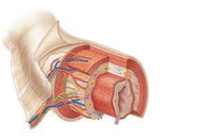
The wall of the alimentary canal consists of four basic layers:
Mucosa: Innermost layer; secretes mucus, digestive enzymes, and hormones; absorbs end products of digestion.
Submucosa: Contains blood vessels, lymphatics, and nerves.
Muscularis externa: Responsible for segmentation and peristalsis; consists of circular and longitudinal muscle layers.
Serosa: Outermost layer; reduces friction as digestive organs move.
The Mouth and Associated Structures
Oral Cavity Anatomy
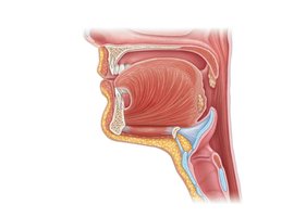
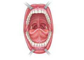
The mouth (oral cavity) is bounded by the lips, cheeks, palate, and tongue. It is lined with stratified squamous epithelium to protect against abrasion. The oral cavity is the entry point for food and the site of initial mechanical and chemical digestion.
The Tongue
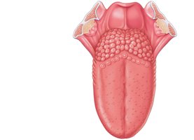
The tongue is a muscular organ that repositions and mixes food during chewing, forms the bolus, and initiates swallowing, speech, and taste. Its surface bears papillae:
Filiform: Provide friction; do not contain taste buds.
Fungiform, Circumvallate, Foliate: Contain taste buds and are involved in gustation.
Salivary Glands
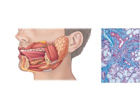
Salivary glands secrete saliva, which cleanses the mouth, moistens food, aids in bolus formation, and contains enzymes (salivary amylase, lingual lipase) that begin the breakdown of starch and fats. Major glands include the parotid, submandibular, and sublingual glands.
Teeth
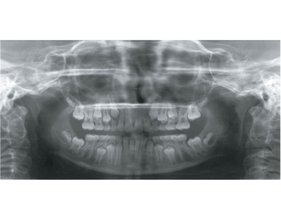
Teeth are essential for mechanical digestion (mastication). Humans have two sets of teeth: 20 deciduous (baby) teeth and 32 permanent teeth. Teeth are classified by shape and function: incisors (cutting), canines (tearing), premolars and molars (grinding).
Pharynx and Esophagus
Pharynx
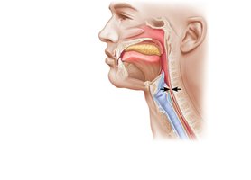
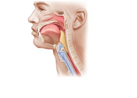
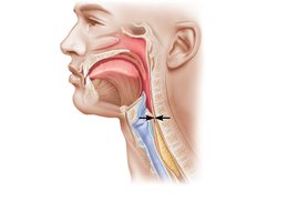
The oropharynx and laryngopharynx allow passage of food, fluids, and air. They are lined with stratified squamous epithelium and contain skeletal muscle layers for swallowing.
Esophagus
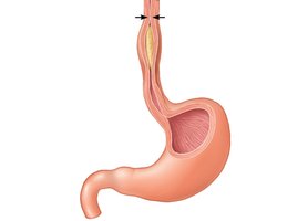
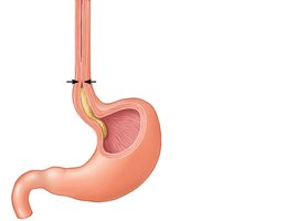
The esophagus is a muscular tube that connects the pharynx to the stomach. It passes through the diaphragm at the esophageal hiatus and joins the stomach at the cardiac orifice.
Stomach
Gross Anatomy and Functions
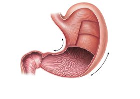
The stomach is a muscular organ with four regions: cardia, fundus, body, and pyloric region. It performs mechanical digestion (churning), chemical digestion (secreting enzymes and acid), and propulsion. Food is converted into chyme before entering the small intestine.
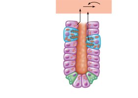
Gastric Gland Secretions
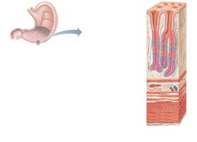
Parietal cells: Secrete hydrochloric acid (HCl) and intrinsic factor (for vitamin B12 absorption).
Chief cells: Secrete pepsinogen, which is activated to pepsin for protein digestion.
Enteroendocrine cells: Secrete hormones such as gastrin, which regulate stomach activity.
Gastric Motility
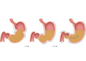
Peristaltic waves mix and propel stomach contents toward the pylorus. The most vigorous mixing occurs near the pyloric region, where small amounts of chyme are released into the duodenum.
Small Intestine
Regions and Functions
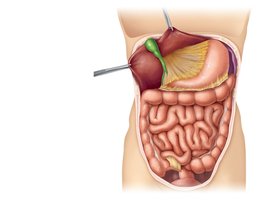
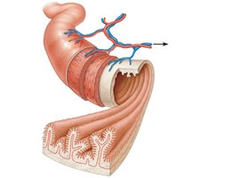
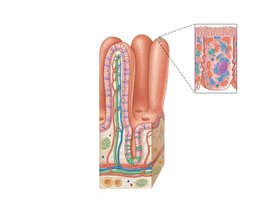
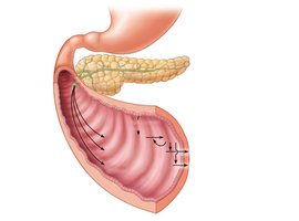
The small intestine is divided into three regions: duodenum, jejunum, and ileum. It is the primary site for chemical digestion and nutrient absorption. Secretions from the liver, gallbladder, and pancreas aid in digestion.
Accessory Digestive Organs
Liver
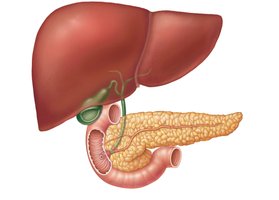
The liver is the largest gland in the body. It processes nutrients, stores vitamins, detoxifies chemicals, and produces bile for fat emulsification.
Gallbladder
The gallbladder stores and concentrates bile, releasing it into the small intestine to aid in fat digestion.
Pancreas
The pancreas has both endocrine (insulin, glucagon) and exocrine (digestive enzymes, bicarbonate) functions. Pancreatic enzymes are crucial for the digestion of proteins, carbohydrates, and fats.
Digestion and Absorption of Nutrients
Protein Digestion
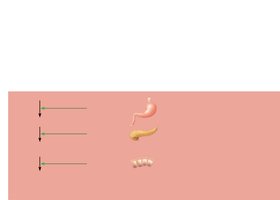
Proteins are digested by pepsin in the stomach and by pancreatic enzymes (trypsin, chymotrypsin, carboxypeptidase) and brush border enzymes in the small intestine. Amino acids are absorbed via cotransport with sodium ions.
Carbohydrate Digestion
Carbohydrates are broken down by salivary and pancreatic amylases and brush border enzymes. Monosaccharides are absorbed by cotransport and facilitated diffusion.
Fat Digestion
Fats are emulsified by bile salts and digested by pancreatic lipases. Fatty acids and monoglycerides are absorbed by diffusion, reassembled into triglycerides, and transported as chylomicrons via the lymphatic system.
Nucleic Acid Digestion
Nucleic acids are digested by pancreatic and brush border enzymes. Their components are absorbed by active transport into the blood.
Large Intestine
Regions and Functions
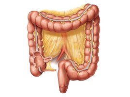
The large intestine includes the cecum, colon (ascending, transverse, descending, sigmoid), rectum, and anal canal. Its main functions are absorption of water, vitamins, and electrolytes, propulsion of feces, and defecation.
Homeostatic Imbalances of the Digestive System
Common Disorders
Gastroesophageal Reflux Disease (GERD): Stomach acid enters the esophagus, causing heartburn and potential damage.
Gastritis: Inflammation of the stomach lining, often due to infection or irritants.
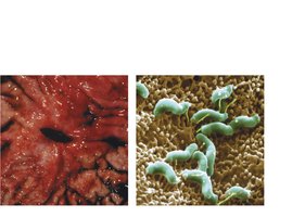
Peptic Ulcers: Erosions of the stomach wall, commonly caused by Helicobacter pylori infection.
Hepatitis: Inflammation of the liver, usually viral in origin.
Cirrhosis: Chronic liver damage leading to scarring and impaired function, often due to alcoholism or hepatitis.
Cancer: Stomach and colon cancers are serious but often asymptomatic in early stages.
Additional info: Regular medical examinations are important for early detection and prevention of digestive system cancers.
