 Back
BackThe Endocrine System: Structure, Function, and Regulation
Study Guide - Smart Notes
 Tailored notes based on your materials, expanded with key definitions, examples, and context.
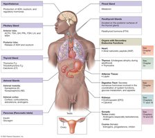
The Endocrine System
Overview of the Endocrine System
The endocrine system is a major regulatory system of the body, working alongside the nervous system to maintain homeostasis. It achieves this by releasing chemical messengers called hormones into the bloodstream, which then act on distant target cells throughout the body.
Homeostasis: Maintained primarily through chemical messages.
Endocrine vs. Nervous System: The nervous system acts quickly with short-lived effects, while the endocrine system acts more slowly but has longer-lasting effects.
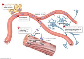
Hormones: Chemical messengers that bind to specific receptors on target cells to elicit a response.
General Features of Hormone Action
Hormones only affect cells that have the appropriate receptor. A single hormone can affect multiple tissues or organs, and a single cell may have receptors for multiple hormones.
Target Cells: Must have specific receptors for a hormone to respond.
Receptor Location: Receptors may be on the plasma membrane or inside the cell.
Classification of Hormones
Hormones are classified based on their chemical structure:
Amino Acid Derivatives: Structurally similar to amino acids (e.g., epinephrine, norepinephrine, thyroid hormones, melatonin).
Peptide Hormones: Chains of amino acids; largest class (e.g., ADH, oxytocin, pituitary hormones).
Lipid Derivatives: Includes steroid hormones (derived from cholesterol) and eicosanoids (derived from fatty acids).
Mechanisms of Hormone Action
Hormones alter cellular operations by changing the types, activities, or quantities of proteins and enzymes within target cells. The mechanism depends on the hormone's solubility:
Plasma Membrane Receptors: Used by hormones that are not lipid-soluble (e.g., peptide hormones, catecholamines). These hormones act as first messengers, activating second messengers inside the cell.
Second Messenger Systems: Small amounts of hormone can activate many second messengers, amplifying the response. Common second messengers include cyclic AMP (cAMP), calcium ions, and cyclic GMP (cGMP).
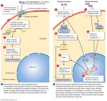
Intracellular Receptors: Used by lipid-soluble hormones (e.g., steroid and thyroid hormones). These hormones cross the plasma membrane, bind to receptors in the cytoplasm or nucleus, and alter gene transcription.
Hormone Secretion and Regulation
Hormones are released into the bloodstream and distributed throughout the body. Their secretion is regulated by negative feedback mechanisms in response to three types of stimuli:
Humoral Stimuli: Changes in the composition of extracellular fluid (e.g., blood calcium levels).
Hormonal Stimuli: Changes in circulating hormone levels (e.g., TSH stimulates thyroid hormone release).
Neural Stimuli: Neural input to endocrine glands (e.g., hypothalamic control of the pituitary gland).
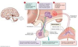
Hypothalamic Control of the Endocrine System
The hypothalamus is the highest level of endocrine control, integrating nervous and endocrine functions. It regulates the pituitary gland through direct neural connections and the secretion of regulatory hormones.
Secretes ADH and oxytocin to the posterior pituitary.
Secretes releasing and inhibiting hormones to control the anterior pituitary.
Controls the adrenal medulla via sympathetic innervation.
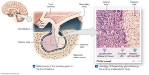
The Pituitary Gland
Anatomy and Structure
The pituitary gland (hypophysis) is located in the sella turcica of the sphenoid bone and is connected to the hypothalamus by the infundibulum. It consists of two distinct lobes: anterior and posterior.
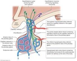
The Hypophyseal Portal System
The anterior lobe of the pituitary gland is regulated by the hypothalamus via the hypophyseal portal system, a network of blood vessels that allows regulatory hormones to reach the anterior pituitary directly.
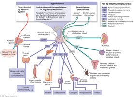
Hormones of the Anterior Pituitary
The anterior pituitary secretes seven major hormones, most of which are tropic (target other endocrine glands):
Thyroid-Stimulating Hormone (TSH): Stimulates thyroid hormone release.
Adrenocorticotropic Hormone (ACTH): Stimulates glucocorticoid secretion from the adrenal cortex.
Follicle-Stimulating Hormone (FSH): Promotes gamete production in ovaries and testes.
Luteinizing Hormone (LH): Triggers ovulation and sex hormone production.
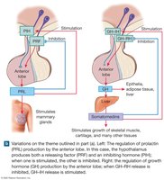
Prolactin (PRL): Stimulates mammary gland development and milk production.
Growth Hormone (GH): Stimulates growth and metabolism in most tissues.
Melanocyte-Stimulating Hormone (MSH): Increases melanin production in the skin (mainly in fetal development).
Hormones of the Posterior Pituitary
The posterior pituitary stores and releases two hormones produced by the hypothalamus:
Antidiuretic Hormone (ADH): Promotes water reabsorption in the kidneys and increases blood pressure.
Oxytocin (OXT): Stimulates uterine contractions during labor and milk ejection during breastfeeding.
The Thyroid and Parathyroid Glands
The Thyroid Gland
The thyroid gland is located anterior to the trachea and inferior to the larynx. It consists of two lobes connected by an isthmus and contains many spherical follicles filled with colloid.
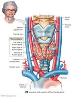
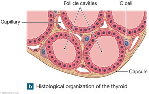
Follicular Cells: Produce thyroid hormones (T3 and T4) in response to TSH.
Thyroxine (T4): Contains four iodine atoms; less potent but more abundant.
Triiodothyronine (T3): Contains three iodine atoms; more potent.
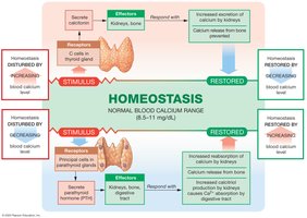
C Cells (Parafollicular Cells): Produce calcitonin, which lowers blood calcium levels.
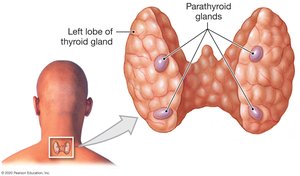
The Parathyroid Glands
The parathyroid glands are small glands embedded in the posterior surface of the thyroid. They secrete parathyroid hormone (PTH), which increases blood calcium levels by stimulating osteoclasts, reducing calcium excretion by the kidneys, and increasing intestinal absorption of calcium.
The Adrenal Glands
Structure and Regions
The adrenal (suprarenal) glands are located on top of each kidney and consist of two regions: the cortex (outer) and the medulla (inner).
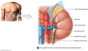
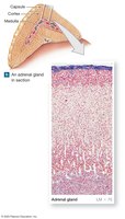
Adrenal Cortex: Produces corticosteroids, including mineralocorticoids (aldosterone), glucocorticoids (cortisol), and androgens.
Adrenal Medulla: Produces catecholamines (epinephrine and norepinephrine) in response to sympathetic stimulation.
The Pineal Gland
Structure and Function
The pineal gland is located in the third ventricle of the brain and produces melatonin, a hormone involved in regulating circadian rhythms and possibly reproductive function.
Melatonin: Secretion is inhibited by light and stimulated by darkness.
Functions: Antioxidant activity, regulation of sleep-wake cycles, and possible effects on puberty.
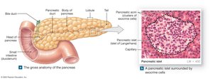
The Endocrine Pancreas
Anatomy and Cell Types
The pancreas contains both exocrine and endocrine cells. The endocrine portion consists of the islets of Langerhans, which contain alpha cells (secrete glucagon) and beta cells (secrete insulin).
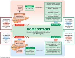
Regulation of Blood Glucose
Insulin: Released in response to high blood glucose; promotes glucose uptake and storage, lowering blood glucose levels.
Glucagon: Released in response to low blood glucose; stimulates glycogen breakdown and glucose release, raising blood glucose levels.
Diabetes Mellitus
Diabetes mellitus is a disorder of glucose metabolism resulting in hyperglycemia. There are two main types:
Type 1 Diabetes: Inadequate insulin production.
Type 2 Diabetes: Insulin resistance in target tissues.
Long-term Effects: Nerve damage, impaired blood flow, retinal damage, and increased risk of heart attack.
Summary Table: Major Endocrine Glands and Their Hormones
Gland | Hormone(s) | Main Function(s) |
|---|---|---|
Pituitary (anterior) | TSH, ACTH, FSH, LH, PRL, GH, MSH | Regulate other endocrine glands, growth, lactation, pigmentation |
Pituitary (posterior) | ADH, OXT | Water balance, uterine contraction, milk ejection |
Thyroid | T3, T4, Calcitonin | Metabolism, calcium regulation |
Parathyroid | PTH | Calcium regulation |
Adrenal cortex | Aldosterone, Cortisol, Androgens | Electrolyte balance, stress response, sex characteristics |
Adrenal medulla | Epinephrine, Norepinephrine | Fight-or-flight response |
Pineal | Melatonin | Circadian rhythms |
Pancreas | Insulin, Glucagon | Blood glucose regulation |
