 Back
BackThe Endocrine System: Structure, Function, and Hormonal Regulation
Study Guide - Smart Notes
 Tailored notes based on your materials, expanded with key definitions, examples, and context.
The Endocrine System
Overview of the Endocrine System
The endocrine system is composed of glands and tissues that secrete hormones, which are chemical messengers responsible for regulating various physiological processes. Unlike exocrine glands, endocrine glands release hormones directly into the bloodstream or interstitial fluid, allowing them to reach distant target organs and tissues.
Endocrine cells: Specialized glandular secretory cells that produce hormones.
Hormones: Chemical messengers that stimulate specific cells or tissues (targets) into action.
Hormones travel through the bloodstream to reach their target cells.
Chemical Classes of Hormones
Amino acid derivatives: Structurally similar to amino acids (e.g., epinephrine, norepinephrine).
Peptide hormones: Short chains of amino acids (e.g., insulin, growth hormone).
Lipid derivatives:
Eicosanoids: Derived from arachidonic acid.
Steroid hormones: Derived from cholesterol (e.g., cortisol, aldosterone).
Regulation of Endocrine Activity
Humoral stimuli: Changes in the composition of extracellular fluid (e.g., blood glucose levels).
Hormonal stimuli: Arrival or removal of a specific hormone in the bloodstream.
Neural stimuli: Arrival of neurotransmitters at neuroglandular junctions.
Hypothalamus and Pituitary Gland
Structure and Function
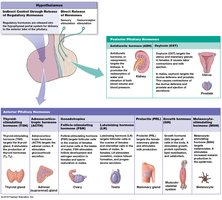
The hypothalamus is the primary regulator of the endocrine system, controlling the activity of the pituitary gland via the infundibulum. The pituitary gland is divided into two lobes: the anterior lobe (glandular tissue) and the posterior lobe (neural tissue).
Hypothalamus: Produces regulatory hormones and two hormones (ADH and oxytocin) stored in the posterior pituitary.
Pituitary gland:
Anterior lobe: Produces and secretes seven hormones.
Posterior lobe: Stores and releases hormones produced by the hypothalamus.
Hypophyseal Portal System
This specialized capillary network connects the hypothalamus to the anterior pituitary, allowing regulatory hormones to reach their target cells efficiently.
Releasing hormones (RH): Stimulate anterior pituitary hormone secretion.
Inhibiting hormones (IH): Inhibit anterior pituitary hormone secretion.
Anterior Pituitary Hormones
Thyroid-stimulating hormone (TSH): Stimulates the thyroid gland to release thyroid hormones.
Adrenocorticotropic hormone (ACTH): Stimulates the adrenal cortex to release glucocorticoids.
Follicle-stimulating hormone (FSH): Stimulates gamete production in ovaries and testes.
Luteinizing hormone (LH): Induces ovulation and secretion of sex hormones.
Prolactin (PRL): Stimulates milk production in mammary glands.
Growth hormone (GH): Promotes protein synthesis and growth in most tissues.
Melanocyte-stimulating hormone (MSH): Increases melanin production in skin.
Posterior Pituitary Hormones
Antidiuretic hormone (ADH): Decreases urine production and constricts blood vessels to raise blood pressure.
Oxytocin: Stimulates uterine contractions during childbirth and milk ejection during breastfeeding.
The Thyroid Gland
Anatomy and Histology
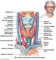
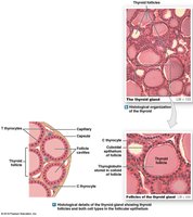
The thyroid gland is located on the anterior surface of the trachea and consists of two lobes connected by an isthmus. Its functional units are thyroid follicles, which are lined by T thyrocytes and contain colloid. C thyrocytes (parafollicular cells) are also present.
Thyroid Hormones
Thyroxine (T4) and Triiodothyronine (T3): Produced by T thyrocytes, stored in colloid, and increase cellular metabolism and oxygen consumption.
Calcitonin: Produced by C thyrocytes, lowers blood calcium levels by inhibiting osteoclast activity and increasing calcium excretion by the kidneys.
Regulation of Thyroid Secretion
TSH from the anterior pituitary stimulates the release of T3 and T4.
Negative feedback mechanisms regulate hormone levels.
The Parathyroid Glands
Structure and Function
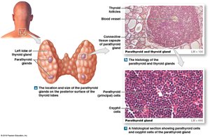
The parathyroid glands are four small glands located on the posterior surface of the thyroid gland. They secrete parathyroid hormone (PTH), which is essential for calcium homeostasis.
Parathyroid Hormone (PTH)
Targets: Bone and kidneys.
Functions: Increases blood calcium levels by stimulating osteoclast activity in bone and enhancing calcium reabsorption in the kidneys.
Summary Table: Thyroid, Parathyroid, and Thymus Hormones
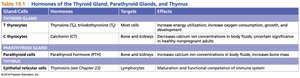
Gland/Cells | Hormones | Targets | Effects |
|---|---|---|---|
T thyrocytes | Thyroxine (T4), Triiodothyronine (T3) | Most cells | Increase energy utilization, oxygen consumption, growth, and development |
C thyrocytes | Calcitonin (CT) | Bone and kidneys | Decrease calcium ion concentrations in body fluids |
Parathyroid cells | Parathyroid hormone (PTH) | Bone and kidneys | Increase calcium ion concentrations in body fluids |
Reticular cells (thymus) | Thymosins | Lymphocytes | Maturation and functional competence of immune system |
The Adrenal Glands
Anatomy and Regions
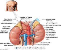
The adrenal glands are located on top of each kidney and consist of two main regions: the cortex and the medulla. Each region produces different types of hormones.
Adrenal Cortex
Zona glomerulosa: Produces mineralocorticoids (e.g., aldosterone) that regulate sodium and potassium balance.
Zona fasciculata: Produces glucocorticoids (e.g., cortisol) that regulate metabolism and stress response.
Zona reticularis: Produces androgens (sex hormones).
Adrenal Medulla
Secretes catecholamines: epinephrine (E) and norepinephrine (NE), which prepare the body for 'fight or flight' responses.
The Pancreas
Pancreatic Islets and Hormones
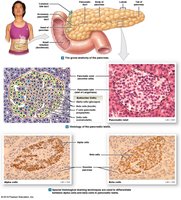
The pancreas contains clusters of endocrine cells called pancreatic islets (islets of Langerhans), which secrete hormones essential for glucose homeostasis.
Major Pancreatic Hormones
Insulin: Produced by beta cells; lowers blood glucose by promoting cellular uptake of glucose.
Glucagon: Produced by alpha cells; raises blood glucose by stimulating glycogen breakdown in the liver.
Somatostatin: Produced by delta cells; inhibits secretion of insulin and glucagon.
Pancreatic polypeptide: Produced by F cells; regulates pancreatic enzyme production and inhibits gallbladder contraction.
Summary Table: Pancreatic Hormones
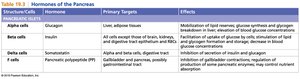
Structure/Cells | Hormone | Primary Targets | Effects |
|---|---|---|---|
Alpha cells | Glucagon | Liver, adipose tissue | Mobilizes lipid reserves, increases blood glucose |
Beta cells | Insulin | Most cells | Facilitates uptake of glucose, lowers blood glucose |
Delta cells | Somatostatin | Alpha and beta cells, digestive tract | Inhibits secretion of insulin and glucagon |
F cells | Pancreatic polypeptide (PP) | Digestive organs | Inhibits gallbladder contraction, regulates enzyme production |
The Hormones of the Reproductive System
Testes and Ovaries
Testes: Produce androgens (e.g., testosterone) that support sperm production and secondary sex characteristics.
Ovaries: Produce estrogens and progestins, which regulate the menstrual cycle, ovulation, and secondary sex characteristics.
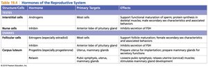
Summary Table: Reproductive Hormones
Structure/Cells | Hormone | Primary Targets | Effects |
|---|---|---|---|
Interstitial cells (testes) | Androgens | Most cells | Support maturation of sperm, protein synthesis in muscles |
Nurse cells (testes) | Inhibin | Anterior pituitary | Inhibits secretion of FSH |
Follicular cells (ovaries) | Estrogens | Most cells | Support follicle maturation, female secondary sex characteristics |
Corpus luteum (ovaries) | Progestins | Uterus, mammary glands | Prepares uterus for implantation, prepares mammary glands for lactation |
Corpus luteum (ovaries) | Relaxin | Pubic symphysis, uterus, mammary glands | Loosens pubic symphysis, relaxes uterine muscles, stimulates mammary gland development |
Additional info:
Hormonal regulation often involves negative feedback loops to maintain homeostasis.
Disorders of the endocrine system can result from hypo- or hypersecretion of hormones, leading to various clinical conditions (e.g., diabetes mellitus, hypothyroidism, Cushing's syndrome).
