 Back
BackThe Endocrine System: Structure, Function, and Disorders
Study Guide - Smart Notes
 Tailored notes based on your materials, expanded with key definitions, examples, and context.
The Endocrine System
Overview of the Endocrine System
The endocrine system is composed of glands and tissues that secrete hormones, which are chemical messengers that regulate various physiological processes throughout the body. These hormones travel through the bloodstream to target cells, which possess specific receptors for each hormone.
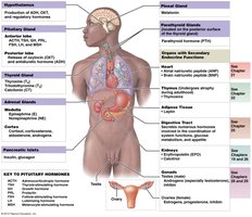
Major Endocrine Organs: Pituitary gland, thyroid gland, parathyroid gland, adrenal gland, pancreas, ovary, and testis.
Hormones: Over 30 different hormones are produced, each with specific regulatory roles.
Target Cells: Cells with receptors that bind and respond to hormones.
Classes of Hormones
Amino Acid Derivatives: Derived from tyrosine (e.g., thyroid hormones, catecholamines) and tryptophan (e.g., serotonin, melatonin).
Peptide Hormones: Chains of amino acids, including glycoproteins (e.g., TSH, LH, FSH), short polypeptides (e.g., ADH, OXT), and proteins (e.g., insulin, GH, prolactin).
Lipid Derivatives: Eicosanoids (from arachidonic acid) and steroid hormones (from cholesterol, e.g., androgens, estrogens, corticosteroids, calcitriol).
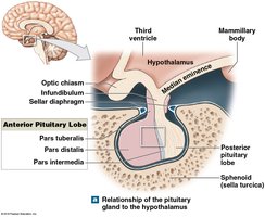
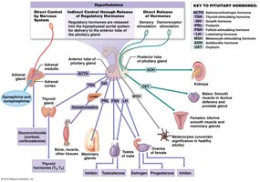
Hypothalamus and Pituitary Gland
Anatomy and Relationship
The pituitary gland (hypophysis) is located within the sella turcica of the sphenoid bone, hanging inferior to the hypothalamus and connected by the infundibulum. It is divided into anterior (adenohypophysis) and posterior (neurohypophysis) lobes, each with distinct functions and hormone production.
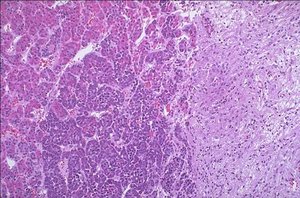
Histology of the Pituitary Gland
The anterior pituitary contains more neuron bodies (granule-like appearance), while the posterior pituitary contains more axons (fiber-like appearance).
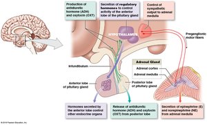
Hypothalamic Control of the Pituitary Gland
The hypothalamus regulates the pituitary gland through three mechanisms:
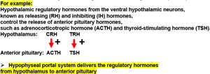
Secretion of regulatory hormones to control the anterior pituitary via the hypophyseal portal system.
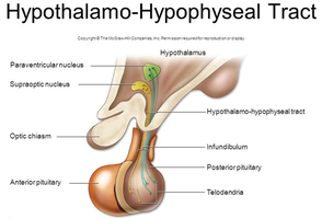
Production of ADH and OXT, transported to the posterior pituitary for release via the hypothalamo-hypophyseal tract.
Direct neural control over the adrenal medulla.
Hypophyseal Portal System
The hypophyseal portal system is a network of blood vessels that links the hypothalamus to the anterior pituitary, allowing regulatory hormones to reach their target cells efficiently.
Releasing Hormones (RH): Stimulate anterior pituitary hormone secretion.
Inhibiting Hormones (IH): Inhibit anterior pituitary hormone secretion.
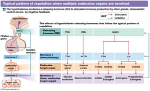
Control of Endocrine Secretion
Endocrine secretion is regulated by positive forward control (stimulation by hypothalamic and pituitary hormones) and negative feedback (inhibition by high levels of target gland hormones).
Hypothalamo-Hypophyseal Tract
This tract consists of axons that transport ADH and OXT from the hypothalamus to the posterior pituitary for release. The supra-optic nucleus produces ADH, and the paraventricular nucleus produces OXT.
Pituitary Gland Hormones, Targets, and Effects
The pituitary gland releases several hormones, each with specific targets and effects. The anterior pituitary produces tropic and general hormones, while the posterior pituitary stores and releases hormones synthesized by the hypothalamus.
Hormone | Stimulus for Release | Target | Effects |
|---|---|---|---|
TSH | Thyrotropin-releasing hormone (TRH) | Thyroid gland | Stimulates thyroid hormone secretion |
ACTH | Corticotropin-releasing hormone (CRH) | Adrenal cortex | Stimulates glucocorticoid secretion |
FSH/LH | Gonadotropin-releasing hormone (GnRH) | Gonads | Stimulates gamete and hormone production |
GH | GHRH/GHIH | Most tissues | Stimulates growth and metabolism |
PRL | PRF/PIH | Mammary glands | Stimulates milk production |
ADH | Neural signals, osmotic pressure | Kidneys | Increases water reabsorption |
OXT | Neural signals | Uterus, mammary glands | Stimulates uterine contractions, milk ejection |
Pituitary Gland Hormone Related Disorders
Growth Hormone Disorders:
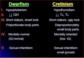
Hyposecretion: Dwarfism
Hypersecretion (childhood): Gigantism
Hypersecretion (adulthood): Acromegaly
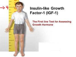
Insulin-like Growth Factor-I (IGF-I) Deficiency: Results in short stature but normal intellectual abilities.
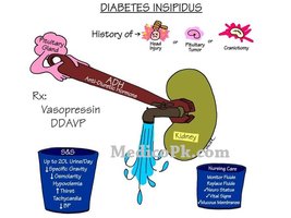
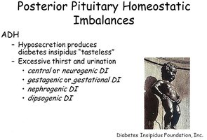
ADH Hyposecretion: Diabetes insipidus, characterized by excessive urination and thirst.


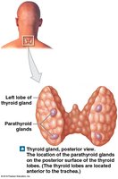
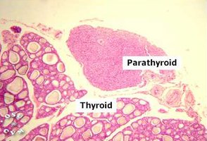
Thyroid Gland
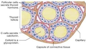
Location, Structure, and Histology
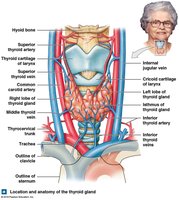
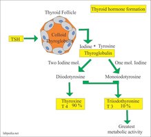
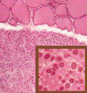
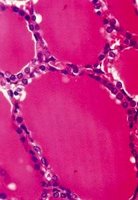
The thyroid gland is located inferior to the thyroid cartilage of the larynx, adjacent to the trachea. It consists of two lobes connected by an isthmus and contains thyroid follicles lined by simple cuboidal epithelium. Follicular cells produce thyroid hormones (T3 and T4), while parafollicular (C) cells produce calcitonin.
Thyroid Hormones
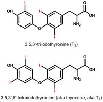
T4 (Thyroxine): Contains four iodine atoms; major form (90%).
T3 (Triiodothyronine): Contains three iodine atoms; active form (10%).
Production: Requires iodine; TSH stimulates synthesis and release.
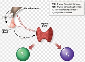
Control of Thyroid Hormone Secretion
Thyroid hormone secretion is regulated by TRH (from hypothalamus) and TSH (from anterior pituitary), with negative feedback from circulating T3 and T4 levels.
Thyroid Gland Disorders
Hyperthyroidism: May cause exophthalmos (protruding eyes).
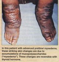
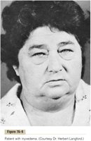
Hypothyroidism: May cause myxedema (swelling due to mucopolysaccharide accumulation).
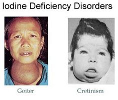
Goiter: Enlarged thyroid gland, often due to iodine deficiency.
Cretinism: Congenital hypothyroidism causing short stature and mental retardation.


Parathyroid Glands
Location, Hormones, and Function
The parathyroid glands are embedded in the posterior surface of the thyroid gland. Principal cells secrete parathyroid hormone (PTH) in response to low blood calcium levels. PTH acts as an antagonist to calcitonin, increasing blood calcium by stimulating osteoclast activity and enhancing calcium reabsorption in the kidneys and intestines.
Thyroid and Parathyroid Gland Hormones, Targets, and Effects
Hormone | Stimulus for Release | Target | Effects |
|---|---|---|---|
Thyroid hormones (T3, T4) | TSH | Most cells | Increase basal metabolic rate, regulate growth and development |
Calcitonin | High blood Ca2+ | Bones | Lowers blood calcium by inhibiting osteoclasts |
PTH | Low blood Ca2+ | Bones, kidneys | Increases blood calcium by stimulating osteoclasts and enhancing reabsorption |

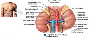
Adrenal Glands
Location, Structure, and Hormones
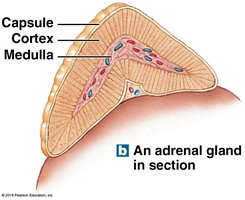
The adrenal glands are located on the superior border of each kidney. Each gland consists of an outer cortex (with three zones) and an inner medulla.
Zona Glomerulosa: Produces mineralocorticoids (e.g., aldosterone).
Zona Fasciculata: Produces glucocorticoids (e.g., cortisol, cortisone).
Zona Reticularis: Produces small quantities of androgens.
Medulla: Produces catecholamines (epinephrine and norepinephrine).
Adrenal Gland Hormone Related Disorders
Pheochromocytoma: Tumor of the adrenal medulla causing excess catecholamine secretion (e.g., increased heart rate and blood pressure).
Cushing's Disease: Hypersecretion of cortisol/cortisone from the cortex, leading to central obesity, moon face, buffalo hump, hypertension, and hyperglycemia.
Addison's Disease: Hyposecretion due to cortex destruction, causing hyperpigmentation, weakness, and tanned skin.
Virilization (Adrenogenital Syndrome): Excess androgen production, especially in females, causing masculinization.
Pancreas
Location, Structure, and Hormones
The pancreas is located retroperitoneally between the stomach and small intestine. It is a mixed gland, with exocrine (digestive) and endocrine (hormonal) functions. The endocrine portion consists of pancreatic islets (Islets of Langerhans), which contain:
Alpha (α) cells: Produce glucagon, which raises blood glucose by stimulating glycogenolysis and gluconeogenesis.
Beta (β) cells: Produce insulin, which lowers blood glucose by stimulating glycogenesis, lipogenesis, and protein synthesis.
Pancreatic Hormone Related Disorders
Diabetes Mellitus: Inadequate insulin production, leading to hyperglycemia, diabetic retinopathy, and diabetic ulcers.
Integration and Modern Advances
The endocrine system integrates with other body systems to maintain homeostasis. Advances in technology and research, including Nobel Prize-winning discoveries, have deepened our understanding of hormone signaling, such as the role of G proteins and second messengers.
Sample Questions and Answers
What tropic hormone stimulates cortisol from the adrenal gland? Answer: Adrenocorticotropic hormone (ACTH)
Under normal conditions, increased levels of thyroid hormone in the blood will cause _______. Answer: A decrease in TSH levels
What hormone also aids the stress response by promoting water retention and acting as a vasoconstrictor? Answer: ADH
When blood glucose levels fall, Answer: Glucagon is released.
