 Back
BackThe Endocrine System: Structure, Function, and Disorders
Study Guide - Smart Notes
 Tailored notes based on your materials, expanded with key definitions, examples, and context.
The Endocrine System: Overview
Introduction to the Endocrine System
The endocrine system works alongside the nervous system to coordinate and integrate the activity of cells throughout the body. It influences metabolic activities by means of hormones transported in the blood. Endocrine responses occur more slowly than nervous responses but tend to last longer. Hormones are produced as needed by various glands and tissues.
Hormones: Chemical messengers that regulate physiological activities and maintain homeostasis.
Some organs, such as the pancreas and gonads, produce both hormones and exocrine products.
The hypothalamus has both neural and endocrine functions.
Other hormone-producing tissues include adipose cells, thymus, small intestine, stomach, kidneys, and heart.
Endocrine vs. Nervous System
Comparison of Control Systems
The endocrine system and nervous system are both control systems but differ in their mechanisms and speed of action.
Nervous system: Uses electrochemical impulses delivered by neurons; responses occur within milliseconds.
Endocrine system: Uses chemical messengers (hormones) carried in the blood; responses are slower but longer-lasting.
Endocrine Glands vs. Exocrine Glands
Types of Glands
There are two main types of glands in the body:
Exocrine glands: Have ducts; secrete non-hormonal products to membrane surfaces (e.g., sweat, sebaceous, mucous, digestive glands).
Endocrine glands: Ductless; release hormones directly into blood or lymph. These glands have rich vascular and lymphatic drainage.
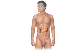
Major endocrine glands include the pituitary, thyroid, parathyroid, adrenal, pineal, and thymus. Some organs (e.g., pancreas, gonads) contain endocrine tissue but are not exclusively endocrine glands.
Chemical Messengers and Hormone Action
Hormones and Their Effects
Hormones are long-distance chemical signals that travel in the blood or lymph to regulate metabolic functions of other cells. They serve to maintain homeostasis by altering the rate of physiological activities in target cells.
Hormones can alter plasma membrane permeability, stimulate protein synthesis, activate or deactivate enzymes, induce secretory activity, and stimulate mitosis.
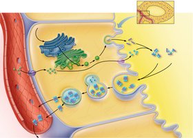
Chemistry of Hormones
Hormones are classified into two main groups:
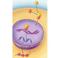
Amino acid-based hormones: Most hormones; water-soluble; act via second-messenger systems and bind to cell surface receptors.
Steroid hormones: Synthesized from cholesterol; lipid-soluble; act via direct gene activation and bind to intracellular receptors.
Second-Messenger Systems
Most amino acid-based hormones use a second-messenger system:
The hormone (first messenger) binds to a receptor on the cell membrane.
This triggers the synthesis of cyclic AMP (cAMP) inside the cell.
cAMP (second messenger) activates enzymes that induce cellular changes.
Intracellular Receptors and Direct Gene Activation
Steroid and thyroid hormones diffuse into target cells and bind to intracellular receptors. The receptor-hormone complex interacts with DNA to initiate transcription and protein synthesis.
Target Cell Specificity and Activation
Target Cell Specificity
Only cells with specific receptors for a hormone will respond to it. The effect of a hormone depends on:
Blood levels of the hormone
Number of receptors on/in the target cell
Affinity (strength) of binding between hormone and receptor
Hormones are inactivated by enzymes or removed by the kidneys and liver.
Control of Hormone Release
Stimuli for Hormone Release
Hormones are released in response to three types of stimuli:
Humoral stimuli: Changes in blood levels of ions/nutrients (e.g., low Ca2+ stimulates PTH release).
Neural stimuli: Nerve fibers stimulate hormone release (e.g., sympathetic stimulation of adrenal medulla).
Hormonal stimuli: Hormones stimulate other endocrine glands (e.g., hypothalamic hormones stimulate pituitary hormones).
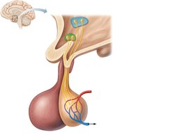
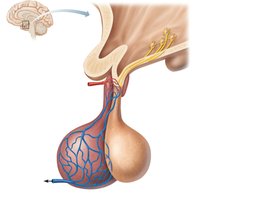
The Pituitary Gland and Hypothalamus
Structure and Function
The pituitary gland (hypophysis) is located in the sella turcica of the sphenoid bone and is attached to the hypothalamus by the infundibulum. It has two major lobes:
Posterior lobe (neurohypophysis): Composed of nervous tissue; stores and releases oxytocin and antidiuretic hormone (ADH) made by the hypothalamus.
Anterior lobe (adenohypophysis): Composed of glandular tissue; manufactures and releases its own hormones in response to hypothalamic stimulation.
Anterior Pituitary Hormones
TSH (Thyroid Stimulating Hormone): Stimulates thyroid hormone secretion.
ACTH (Adrenocorticotropic Hormone): Stimulates adrenal cortex to release corticosteroids.
FSH (Follicle Stimulating Hormone): Stimulates gamete production.
LH (Luteinizing Hormone): Promotes production of estrogen and testosterone.
GH (Growth Hormone): Stimulates growth, especially in bones and muscles; promotes protein synthesis and fat utilization.
Prolactin: Stimulates milk production.
Posterior Pituitary Hormones
ADH (Antidiuretic Hormone): Promotes water reabsorption in kidneys; inhibited by alcohol.
Oxytocin: Stimulates uterine contractions during childbirth and milk ejection during breastfeeding.
Thyroid and Parathyroid Glands
Thyroid Gland Structure and Function
The thyroid gland is located in the anterior neck and consists of two lobes connected by an isthmus. It produces thyroid hormone (TH) and calcitonin.
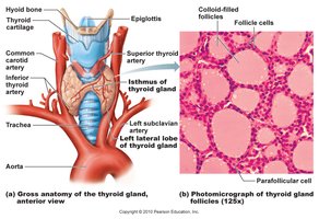
Thyroid Hormone (TH)
Major metabolic hormone; affects nearly every cell in the body.
Composed of T4 (thyroxine) and T3 (triiodothyronine).
Increases basal metabolic rate, heat production, and regulates tissue growth.
Calcitonin
Produced by parafollicular (C) cells of the thyroid.
Released in response to high blood calcium; lowers blood calcium by stimulating bone uptake.
Parathyroid Glands and Hormone
The parathyroid glands are small glands on the posterior thyroid. They secrete parathyroid hormone (PTH), the main regulator of blood calcium.
PTH increases blood calcium by stimulating bone resorption, increasing kidney reabsorption, and enhancing intestinal absorption (via vitamin D activation).
Adrenal (Suprarenal) Glands
Structure and Hormones
The adrenal glands sit atop the kidneys and consist of two regions:
Adrenal cortex: Produces corticosteroids (mineralocorticoids, glucocorticoids, gonadocorticoids).
Adrenal medulla: Produces catecholamines (epinephrine and norepinephrine).
Adrenal Cortex Hormones
Mineralocorticoids (e.g., aldosterone): Regulate Na+ and K+ balance; affect blood pressure.
Glucocorticoids (e.g., cortisol): Influence metabolism, help resist stress, maintain blood glucose and pressure.
Gonadocorticoids: Sex hormones; contribute to puberty and secondary sex characteristics.
Adrenal Medulla Hormones
Secretes epinephrine and norepinephrine in response to stress; increases heart rate, blood pressure, and blood glucose.
Pineal Gland
Melatonin and Circadian Rhythms
The pineal gland secretes melatonin, which regulates sleep-wake cycles and other rhythmic physiological processes.
Pancreas
Structure and Function
The pancreas has both exocrine (digestive enzyme production) and endocrine (hormone production) functions. The islets of Langerhans contain alpha cells (produce glucagon) and beta cells (produce insulin).
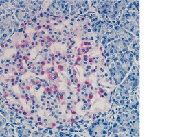
Glucagon and Insulin
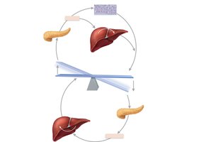
Glucagon: Released when blood glucose is low; stimulates glycogen breakdown and glucose synthesis in the liver.
Insulin: Released when blood glucose is high; promotes glucose uptake by cells, glycogen formation, and fat storage.
Gonads
Sex Hormones
Ovaries: Produce estrogens and progesterone; regulate female reproductive development and function.
Testes: Produce testosterone; regulate male reproductive development and function.
Thymus
Immune Function
The thymus is involved in the development of T lymphocytes and the immune response, especially during youth.
Selected Endocrine Disorders
Growth Hormone Disorders
Gigantism: Hypersecretion of GH in children; excessive growth.
Acromegaly: Hypersecretion of GH in adults; overgrowth of hands, feet, face.
Pituitary dwarfism: Hyposecretion of GH in children; short stature.
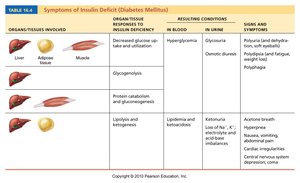
Diabetes Insipidus and Mellitus
Diabetes insipidus: ADH deficiency; excessive urine output and thirst.
Diabetes mellitus (DM): Insulin deficiency or inactivity; high blood glucose, polyuria, polydipsia, polyphagia.
Other Disorders
Myxedema: Hypothyroidism; low metabolic rate, edema.
Graves' disease: Hyperthyroidism; elevated metabolism, weight loss.
Cushing's syndrome: Excess glucocorticoids; high blood glucose, muscle/bone loss.
Addison's disease: Deficits in glucocorticoids/mineralocorticoids; weight loss, dehydration, low blood glucose.
