Back
The Endocrine System: Structure, Function, and Clinical Relevance
Study Guide - Smart Notes
 Tailored notes based on your materials, expanded with key definitions, examples, and context.
The Endocrine System
Overview of the Endocrine System
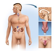
The endocrine system is a collection of glands that secrete hormones directly into the bloodstream to regulate various body functions. These hormones control processes such as growth, metabolism, and reproduction. The major organs of the endocrine system include the pineal gland, pituitary gland, hypothalamus, thyroid gland, thymus, adrenal glands, pancreas, and gonads (ovaries and testes).
Hormones: Chemical messengers that influence target cells and organs.
Endocrine glands: Ductless glands that release hormones into the blood.
Functions: Regulation of metabolism, growth, development, tissue function, and mood.
Pineal Gland
Structure and Function
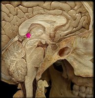
The pineal gland is a small endocrine gland located dorsal to the thalamus in the brain. It is composed mainly of pinealocytes, which secrete the hormone melatonin, derived from serotonin.
Melatonin: Regulates sleep-wake cycles (circadian rhythms) and is involved in the timing of other biological rhythms such as appetite and body temperature.
Involution: The pineal gland shrinks after age 7, decreasing by about 75% by the end of puberty.
Thymus
Structure and Function
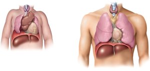
The thymus is located in the upper thoracic cavity, anterior to the heart. It is larger in infants and children and shrinks after puberty (involution). The thymus secretes thymosins, hormones that stimulate the maturation of T lymphocytes, which are crucial for immune defense.
Thymosins: Promote development and differentiation of T cells.
Involution: The thymus decreases to about 10% of its original size in adulthood.
Thyroid Gland
Structure and Histology
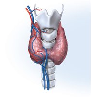
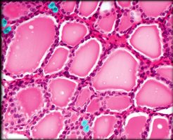
The thyroid gland is the largest endocrine gland, located in the anterior neck, inferior to the larynx. It consists of thyroid follicles filled with protein-rich colloid and surrounded by follicular cells.
Follicular cells: Secrete thyroid hormones (T3 and T4).
Parafollicular (C) cells: Secrete calcitonin.
Thyroid Hormones: Synthesis, Storage, and Secretion
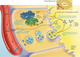
Thyroid hormones are synthesized from the precursor thyroglobulin and require dietary iodine. The main hormones are thyroxine (T4) and triiodothyronine (T3).
Synthesis: Involves iodination of tyrosine residues in thyroglobulin to form MIT (monoiodotyrosine) and DIT (diiodotyrosine).
Hormone formation: MIT + DIT = T3; DIT + DIT = T4 (more abundant).
Secretion: T3 and T4 are released into the bloodstream.
Regulation of Thyroid Hormone Secretion
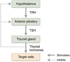
Thyroid hormone secretion is regulated by the hypothalamic-pituitary-thyroid axis. Thyrotropin-releasing hormone (TRH) from the hypothalamus stimulates the anterior pituitary to release thyroid-stimulating hormone (TSH), which in turn stimulates the thyroid gland.
Negative feedback: High levels of thyroid hormones inhibit TRH and TSH secretion.
Major Effects of Thyroid Hormones
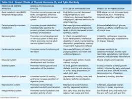
Thyroid hormones increase metabolic rate, oxygen consumption, and heat production (calorigenic effect). They also enhance growth hormone secretion, alertness, and reflexes.
Systems affected: Cardiovascular, nervous, muscular, skeletal, gastrointestinal, reproductive, and integumentary systems.
System | Normal Effect | Hyposecretion | Hypersecretion |
|---|---|---|---|
Metabolism | Increases BMR, heat production | Low BMR, cold intolerance | High BMR, heat intolerance |
Nervous | Promotes development | Mental sluggishness | Restlessness, insomnia |
Cardiovascular | Increases heart rate | Decreased function | Rapid heart rate |
Clinical Imbalances of Thyroid Function
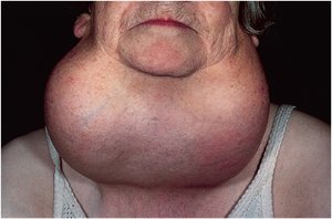
Hyposecretion (Myxedema): Low metabolic rate, feeling chilled, mental sluggishness, lethargy. If due to iodine deficiency, a goiter may develop.
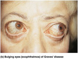
Hypersecretion (Graves' Disease): Autoimmune disorder where antibodies mimic TSH, causing excessive thyroid hormone release. Symptoms include elevated metabolic rate, sweating, nervousness, and exophthalmos (bulging eyes).
Calcitonin
Calcitonin is produced by parafollicular (C) cells in response to high blood calcium levels. It lowers blood calcium by inhibiting osteoclast activity and increasing calcium excretion in the kidneys. It acts as an antagonist to parathyroid hormone (PTH).
Parathyroid Glands
Structure and Function
The parathyroid glands are small glands located on the posterior surface of the thyroid gland. They secrete parathyroid hormone (PTH), which increases blood calcium levels by stimulating osteoclasts, increasing intestinal absorption, and reducing urinary excretion of calcium.
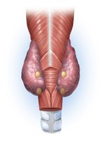
Parathyroid Hormone (PTH)
Bone response: Stimulates osteoclasts to release calcium from bone.
Intestinal response: Increases absorption of calcium via increased calcitriol synthesis.
Kidney response: Reduces calcium loss in urine.
Adrenal (Suprarenal) Glands
Structure and Function
The adrenal glands are located on top of the kidneys and consist of two regions: the cortex and the medulla.
Adrenal cortex: Produces corticosteroids (mineralocorticoids, glucocorticoids, sex steroids).
Adrenal medulla: Produces catecholamines (epinephrine and norepinephrine).
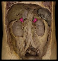
Adrenal Cortex Hormones
Mineralocorticoids (e.g., aldosterone): Regulate electrolyte and water balance by stimulating sodium retention and potassium excretion.
Glucocorticoids (e.g., cortisol): Promote fat and protein breakdown, gluconeogenesis, and help the body adapt to stress. Long-term use can suppress the immune system.
Sex steroids (e.g., androgens): Influence libido and development of secondary sex characteristics.
Adrenal Medulla Hormones
Epinephrine and norepinephrine: Mediate the fight-or-flight response as part of the sympathetic nervous system.
Pancreas
Structure and Function
The pancreas has both exocrine and endocrine functions. The endocrine portion consists of pancreatic islets (islets of Langerhans), which contain alpha, beta, and delta cells.
Alpha (A) cells: Secrete glucagon, which raises blood glucose by promoting glycogen breakdown and gluconeogenesis in the liver.
Beta (B) cells: Secrete insulin, which lowers blood glucose by promoting cellular uptake of glucose and synthesis of glycogen, fat, and protein.
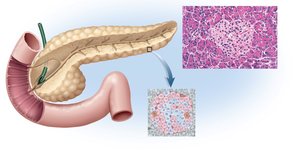
Diabetes Mellitus
Diabetes mellitus is the most prevalent metabolic disease, characterized by low secretion or inaction of insulin. Symptoms include polyuria (excess urine), polydipsia (thirst), polyphagia (hunger), and elevated blood and urine glucose.
Type 1 diabetes: Autoimmune destruction of beta cells; requires insulin therapy.
Type 2 diabetes: Insulin resistance; associated with obesity, heredity, and certain ethnicities. Managed with diet, exercise, and sometimes medication.
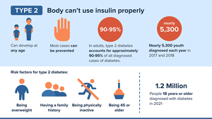
Gonads (Ovaries and Testes)
Structure and Function
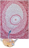
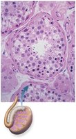
The gonads are both endocrine and exocrine organs. The ovaries produce eggs and secrete estrogen and progesterone, while the testes produce sperm and secrete testosterone.
Ovaries: Estrogen from granulosa cells supports development of the female reproductive system, pregnancy, and lactation. The corpus luteum secretes progesterone after ovulation.
Testes: Testosterone from interstitial cells supports development of the male reproductive system, sex drive, and sperm production.