 Back
The Endocrine System: Structure, Function, and Regulation
Study Guide - Smart Notes
 Tailored notes based on your materials, expanded with key definitions, examples, and context.
The Endocrine System
Overview of the Endocrine System
The endocrine system is a network of glands that produce and secrete hormones to regulate various bodily functions, including metabolism, growth, reproduction, and homeostasis. Hormones are chemical messengers that travel through the bloodstream to target organs and tissues.
Key Glands: Pituitary, thyroid, parathyroid, adrenal, pancreas, gonads, pineal, and others.
Hormones: Include peptide, steroid, and amino acid-based hormones.
Regulation: Often involves negative feedback mechanisms to maintain homeostasis.
The Posterior Pituitary
Hormones of the Posterior Pituitary
The posterior pituitary stores and releases two important hormones produced by the hypothalamus: antidiuretic hormone (ADH) and oxytocin.
ADH (Antidiuretic Hormone): Regulates water balance by inhibiting urine formation. Released in response to increased blood solute concentration.
Oxytocin: Stimulates uterine contractions during childbirth and triggers milk ejection in lactating women. Also involved in sexual arousal and orgasm.
Mechanism: Both hormones use the PIP-calcium second-messenger system at their targets.
Homeostatic Imbalances of ADH
ADH Deficiency: Causes diabetes insipidus, characterized by excessive urine output and intense thirst.
ADH Hypersecretion: Can result from neurosurgery, trauma, or cancer, leading to syndrome of inappropriate ADH secretion (SIADH).
Thyroid Gland
Anatomy and Histology of the Thyroid Gland
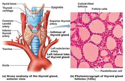
The thyroid gland consists of two lateral lobes connected by an isthmus and is composed of follicles that produce thyroglobulin. Parafollicular cells produce calcitonin.
Follicles: Filled with colloid (thyroglobulin + iodine), precursor to thyroid hormone.
Parafollicular Cells: Produce calcitonin, a hormone involved in calcium regulation.
Thyroid Hormone (TH)
Thyroid hormone consists of two related compounds: T4 (thyroxine) and T3 (triiodothyronine).
T4: Contains two tyrosine molecules and four iodine atoms.
T3: Contains two tyrosines and three iodine atoms; more active than T4.
Functions: Increases metabolic rate, heat production, maintains blood pressure, regulates tissue growth, and is essential for skeletal, nervous, and reproductive system development.
Synthesis of Thyroid Hormone
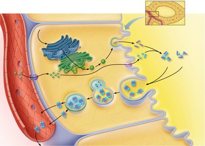
The synthesis of thyroid hormone involves several steps, including the uptake of iodide, oxidation to iodine, attachment to tyrosine, and formation of T3 and T4.
Thyroglobulin: Synthesized and discharged into the follicle lumen.
Iodide: Actively transported into cells, oxidized to iodine, and attached to tyrosine.
Formation: Iodinated tyrosines link to form T3 and T4, which are then released into the bloodstream.
Transport and Regulation of Thyroid Hormone
Transport: T4 and T3 are carried by thyroxine-binding globulins (TBGs).
Activity: T3 is ten times more active than T4; peripheral tissues convert T4 to T3.
Regulation: Negative feedback inhibits TSH release; hypothalamic TRH can override feedback during pregnancy or cold exposure.
Homeostatic Imbalances of Thyroid Hormone
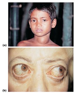
Hyposecretion in Adults: Myxedema (mucous swelling); endemic goiter if due to iodine deficiency.
Hyposecretion in Infants: Cretinism.
Hypersecretion: Graves’ disease, an autoimmune disorder causing continuous stimulation of TH release.
Calcitonin
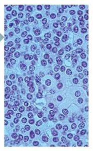
Calcitonin is produced by parafollicular (C) cells and acts as an antagonist to parathyroid hormone (PTH).
Function: Inhibits osteoclast activity, stimulates Ca2+ uptake into bone matrix.
Regulation: Controlled by blood Ca2+ concentration via negative feedback.
Clinical Note: Removal of thyroid does not affect Ca2+ homeostasis in humans.
Parathyroid Glands
Anatomy and Histology of Parathyroid Glands
The parathyroid glands are small glands located on the posterior aspect of the thyroid. They contain chief cells (secrete PTH) and oxyphil cells (function unknown).
PTH (Parathyroid Hormone): Most important hormone in Ca2+ homeostasis.
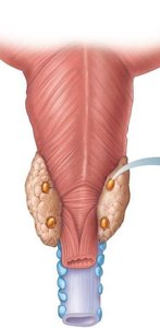
Functions of Parathyroid Hormone
Bone: Stimulates osteoclasts to digest bone matrix, releasing Ca2+ and phosphate.
Kidneys: Enhances reabsorption of Ca2+, promotes activation of vitamin D, increases Ca2+ absorption from intestine.
Regulation: Negative feedback; rising blood Ca2+ inhibits PTH release.
Homeostatic Imbalances of PTH
Hyperparathyroidism: Bones soften and deform, elevated Ca2+ depresses nervous system, kidney stones may form.
Hypoparathyroidism: Results in tetany, respiratory paralysis, and death.
Adrenal (Suprarenal) Glands
Anatomy and Histology of Adrenal Glands
The adrenal glands are paired, pyramid-shaped organs atop the kidneys, consisting of an adrenal cortex and medulla.
Adrenal Cortex: Three layers producing corticosteroids: zona glomerulosa (mineralocorticoids), zona fasciculata (glucocorticoids), zona reticularis (gonadocorticoids).
Adrenal Medulla: Nervous tissue, part of the sympathetic nervous system.
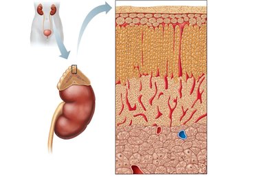
Mineralocorticoids (Aldosterone)
Function: Regulate electrolytes (Na+, K+) in extracellular fluid.
Aldosterone: Most potent mineralocorticoid; stimulates Na+ reabsorption and water retention by kidneys.
Regulation: Renin-angiotensin mechanism, plasma K+ concentration, ACTH, and atrial natriuretic peptide (ANP).
Homeostatic Imbalances of Aldosterone
Aldosteronism: Hypersecretion due to adrenal tumors; causes hypertension, edema, and abnormal neuron/muscle function.
Glucocorticoids (Cortisol)
Function: Maintain blood sugar levels, blood pressure, and promote gluconeogenesis (formation of glucose from fats and proteins).
Cortisol: Most significant glucocorticoid; released in response to ACTH, stress, and activity patterns.
Homeostatic Imbalances of Glucocorticoids
Hypersecretion: Cushing’s syndrome; depresses cartilage/bone formation, immune system, and causes cardiovascular, neural, and GI changes.
Hyposecretion: Addison’s disease; deficits in mineralocorticoids, decreased glucose and Na+, weight loss, dehydration, hypotension.
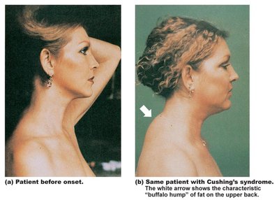
Gonadocorticoids (Sex Hormones)
Androgens: Converted to testosterone or estrogens; contribute to puberty, secondary sex characteristics, and sex drive.
Adrenal Medulla
Chromaffin Cells: Secrete epinephrine (80%) and norepinephrine (20%).
Effects: Increase blood glucose, constrict blood vessels, increase heart rate, divert blood to brain, heart, and skeletal muscle.
Epinephrine: Stimulates metabolic activities, bronchial dilation, blood flow to muscles and heart.
Norepinephrine: Influences peripheral vasoconstriction and blood pressure.
Pineal Gland
Structure and Function
The pineal gland is a small gland that secretes melatonin, which regulates day/night cycles and physiological rhythms.
Melatonin: May affect timing of puberty, sleep, appetite, and body temperature.
Pancreas
Anatomy and Histology of the Pancreas
The pancreas is a triangular gland with both exocrine and endocrine functions.
Acinar Cells: Exocrine cells producing digestive enzymes.
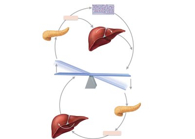
Pancreatic Islets (Islets of Langerhans): Endocrine cells; alpha cells produce glucagon, beta cells produce insulin.
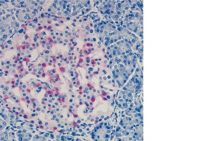
Glucagon
Major Target: Liver; promotes glycogenolysis (breakdown of glycogen), gluconeogenesis (synthesis of glucose), and release of glucose into blood.
Insulin
Function: Lowers blood glucose, enhances glucose uptake by fat and muscle cells, inhibits glycogenolysis and gluconeogenesis.
Action: Activates tyrosine kinase receptor, increases glucose uptake, catalyzes glucose oxidation, glycogen formation, and conversion to fat.
Homeostatic Imbalances of Insulin
Diabetes Mellitus (DM): Due to hyposecretion or hypoactivity of insulin; characterized by polyuria, polydipsia, and polyphagia.
Hyperinsulinism: Excessive insulin secretion; results in hypoglycemia, disorientation, unconsciousness.
Gonads and Placenta
Ovaries and Placenta
Ovaries: Produce estrogens and progesterone; responsible for maturation of female reproductive organs, secondary sexual characteristics, breast development, and uterine cycle.
Placenta: Secretes estrogens, progesterone, and human chorionic gonadotropin (hCG).
Testes
Testes: Produce testosterone; initiates maturation of male reproductive organs, secondary sexual characteristics, sex drive, sperm production, and maintenance of reproductive organs.
Other Hormone-Producing Structures
Additional Endocrine Functions
Several other organs produce hormones that regulate various physiological processes.
Heart: Atrial natriuretic peptide (ANP) reduces blood pressure, blood volume, and Na+ concentration.
Gastrointestinal Tract: Enteroendocrine cells produce gastrin, secretin, and cholecystokinin.
Kidneys: Erythropoietin stimulates red blood cell production; renin initiates the renin-angiotensin mechanism.
Skin: Produces cholecalciferol, precursor to vitamin D.
Adipose Tissue: Leptin regulates appetite and energy expenditure.
Skeleton: Osteocalcin stimulates insulin secretion and improves glucose handling.
Thymus: Produces thymulin, thymopoietins, and thymosins for T lymphocyte development.
Developmental Aspects of the Endocrine System
Effects of Aging and Environmental Factors
Pollutants: Exposure to chemicals can disrupt hormone function, especially sex hormones, thyroid hormone, and glucocorticoids.
Aging: Ovaries become unresponsive to gonadotropins, estrogen deficiency increases bone vulnerability; testosterone declines with age; GH and TH levels decrease, leading to muscle atrophy and lower metabolic rates; PTH remains constant but bone effects increase in older women.
Key Equations and Concepts
Hormone Regulation by Negative Feedback
General Feedback Equation:
Gluconeogenesis (Cortisol Effect)
Equation:
Blood Glucose Regulation (Insulin and Glucagon)
Equation:
Summary Table: Major Endocrine Glands and Hormones
Gland | Hormone(s) | Main Function |
|---|---|---|
Pituitary (Posterior) | ADH, Oxytocin | Water balance, uterine contraction, milk ejection |
Thyroid | T3, T4, Calcitonin | Metabolism, calcium regulation |
Parathyroid | PTH | Calcium homeostasis |
Adrenal Cortex | Aldosterone, Cortisol, Androgens | Electrolyte balance, stress response, sex characteristics |
Adrenal Medulla | Epinephrine, Norepinephrine | Fight-or-flight response |
Pancreas | Insulin, Glucagon | Blood glucose regulation |
Gonads | Estrogen, Progesterone, Testosterone | Reproduction, secondary sex characteristics |
