 Back
The Endocrine System: Structure, Function, and Hormonal Regulation
Study Guide - Smart Notes
 Tailored notes based on your materials, expanded with key definitions, examples, and context.
The Endocrine System: Overview
General Function and Organization
The endocrine system works in concert with the nervous system to coordinate and integrate the activity of body cells. It influences metabolic activities by means of hormones transported in the blood. Endocrine responses are generally slower but longer-lasting than nervous system responses, which rely on action potentials.
Hormones are chemical messengers that regulate processes such as growth, metabolism, and reproduction.
Some organs, such as the pancreas and gonads, have both endocrine (hormone-secreting, ductless) and exocrine (duct-secreting) functions.
The hypothalamus is a neuroendocrine organ, integrating neural and hormonal signals.
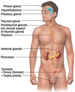
Chemical Messengers of the Endocrine System
Types of Chemical Messengers
Hormones are long-distance chemical signals that travel in the blood or lymph and regulate various physiological processes. Other local chemical messengers include autocrines and paracrines, but these are not considered part of the endocrine system.
Autocrines: Act on the same cells that secrete them (e.g., prostaglandins).
Paracrines: Affect nearby cells (e.g., histamine, serotonin).
Chemistry of Hormones
Hormone Classes
Hormones are classified based on their chemical structure, which determines their mechanism of action.
Amino acid-based hormones: Include amines, peptides, and proteins.
Steroid hormones: Synthesized from cholesterol; include gonadal and adrenocortical hormones.
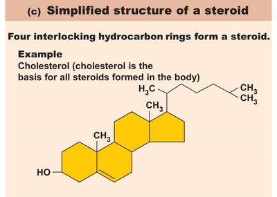
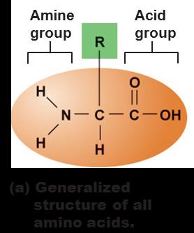
Mechanisms of Hormone Action
How Hormones Affect Target Cells
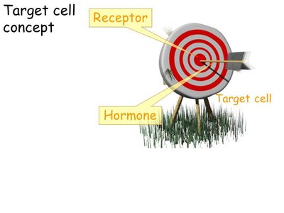
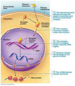
Hormones act on target cells that have specific receptors, altering cellular activity in several ways:
Altering membrane permeability or membrane potential by opening/closing ion channels
Stimulating synthesis of proteins or regulatory molecules
Activating or deactivating enzyme systems
Inducing secretory activity
Stimulating mitosis
Water-Soluble vs. Lipid-Soluble Hormones
Water-soluble hormones (all amino acid-based except thyroid hormone): Cannot enter target cells; act on plasma membrane receptors and use second messengers (e.g., cAMP).
Lipid-soluble hormones (steroids and thyroid hormone): Act on intracellular receptors that directly activate genes.
Target Cell Specificity and Activation
Specificity and Regulation
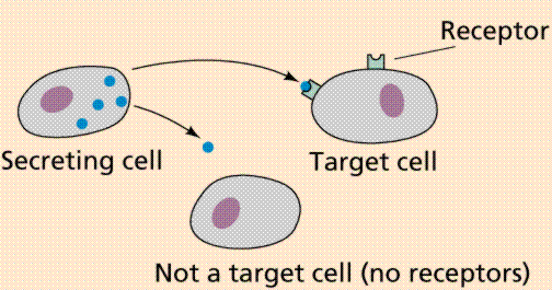
Only target cells with the appropriate receptors respond to a given hormone. The degree of target cell activation depends on:
Number of receptors on/in the target cell
Blood levels of the hormone
Affinity (strength) of binding between hormone and receptor
Regulation of Receptor Number
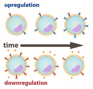
Up-regulation: Target cells form more receptors in response to low hormone levels.
Down-regulation: Target cells lose receptors in response to high hormone levels.
Hormone Levels in the Blood
Transport, Inactivation, and Removal
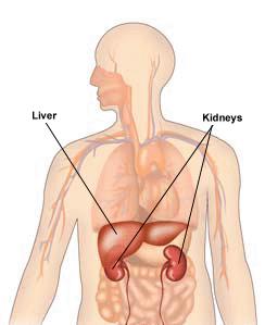
Hormones circulate in the blood either free or bound to plasma proteins. The concentration of a hormone in the blood reflects its rate of release and the speed of inactivation/removal, which occurs via degrading enzymes, kidneys, or liver. The half-life is the time required for a hormone's blood level to decrease by half.
Control of Hormone Release
Feedback Mechanisms and Stimuli
Hormone secretion is primarily regulated by negative feedback mechanisms and is triggered by three types of stimuli:
Humoral stimuli: Changing blood levels of ions/nutrients (e.g., Ca2+ levels regulate PTH release).
Neural stimuli: Nerve fibers stimulate hormone release (e.g., sympathetic stimulation of adrenal medulla).
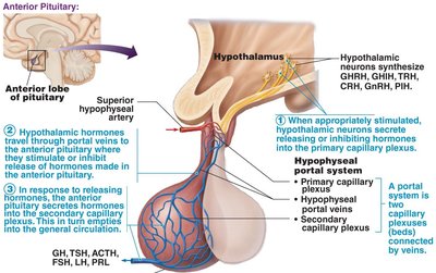
Hormonal stimuli: Hormones stimulate other endocrine glands (e.g., hypothalamic hormones regulate anterior pituitary).
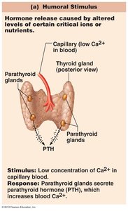
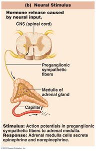
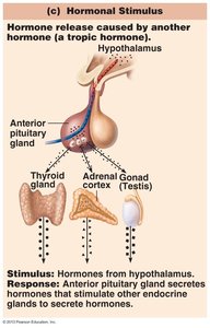
Interaction of Hormones at Target Cells
Types of Hormonal Interactions
Permissiveness: One hormone requires another to exert its full effect (e.g., reproductive hormones and thyroid hormone).
Synergism: More than one hormone produces the same effect, amplifying the response (e.g., glucagon and epinephrine).
Antagonism: One hormone opposes the action of another (e.g., insulin vs. epinephrine on blood glucose).
The Pituitary Gland and Hypothalamus
Structure and Function
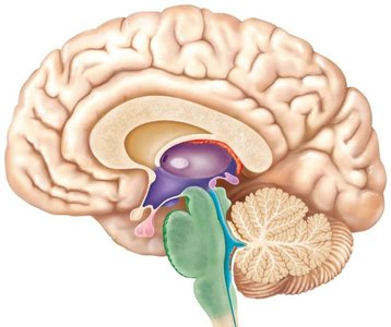
The pituitary gland (hypophysis) consists of two major lobes:
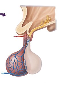
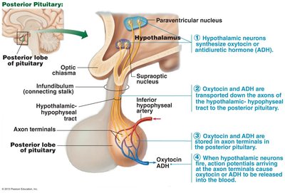
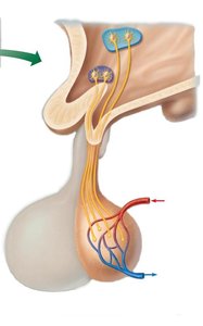
Posterior pituitary: Neural tissue; stores and releases oxytocin and antidiuretic hormone (ADH) produced by the hypothalamus.
Anterior pituitary: Glandular tissue; produces and releases several hormones under hypothalamic regulation.
Anterior Pituitary Hormones
Major Hormones and Their Functions
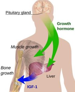
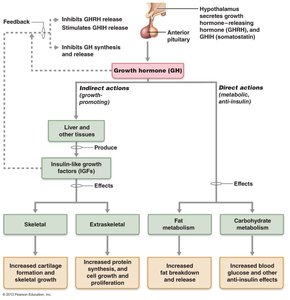
Growth hormone (GH): Stimulates growth of bones and muscles; promotes protein synthesis and fat utilization.
Thyroid-stimulating hormone (TSH): Stimulates thyroid gland activity.
Adrenocorticotropic hormone (ACTH): Stimulates adrenal cortex to release corticosteroids.
Follicle-stimulating hormone (FSH): Stimulates gamete production.
Luteinizing hormone (LH): Promotes production of gonadal hormones.
Prolactin (PRL): Stimulates milk production.
TSH, ACTH, FSH, and LH are tropic hormones, meaning they regulate the activity of other endocrine glands.
Growth Hormone (GH) Regulation and Effects
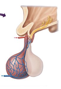
GH release is regulated by growth hormone-releasing hormone (GHRH) and growth hormone-inhibiting hormone (GHIH, somatostatin) from the hypothalamus. Most effects are mediated indirectly by insulin-like growth factors (IGFs).
Homeostatic Imbalances of GH
Hypersecretion: Gigantism in children, acromegaly in adults.
Hyposecretion: Pituitary dwarfism in children.


Thyroid-Stimulating Hormone (TSH)
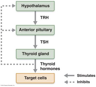
TSH is produced by thyrotrophs of the anterior pituitary and stimulates the thyroid gland. Its release is regulated by thyrotropin-releasing hormone (TRH) and inhibited by rising thyroid hormone levels (negative feedback).
Adrenocorticotropic Hormone (ACTH)
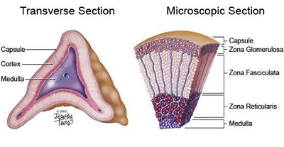
ACTH stimulates the adrenal cortex to release corticosteroids. Its release is triggered by corticotropin-releasing hormone (CRH) and influenced by stress, fever, and hypoglycemia.
Gonadotropins (FSH and LH)
FSH and LH regulate gamete production and gonadal hormone secretion. Their release is triggered by gonadotropin-releasing hormone (GnRH) and suppressed by gonadal hormones (negative feedback).
Prolactin (PRL)
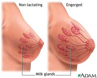
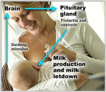
PRL stimulates milk production. Its release is primarily inhibited by prolactin-inhibiting hormone (PIH, dopamine). Suckling increases PRL release, promoting continued milk production.
Posterior Pituitary Hormones
Oxytocin and Antidiuretic Hormone (ADH)
Oxytocin: Stimulates uterine contractions during childbirth and milk ejection during lactation; involved in sexual arousal and orgasm.
ADH: Regulates water balance by increasing water reabsorption in the kidneys; released in response to high blood solute concentration.
Homeostatic Imbalances of ADH
Deficiency: Diabetes insipidus (excessive urine output, thirst).
Hypersecretion: Syndrome of inappropriate ADH secretion (SIADH; fluid retention, low sodium).
Thyroid Gland and Hormones
Structure and Function
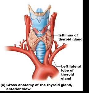
The thyroid gland consists of two lateral lobes connected by an isthmus. Follicle cells produce thyroglobulin, and parafollicular cells produce calcitonin. The colloid (thyroglobulin + iodine) is the precursor of thyroid hormone.
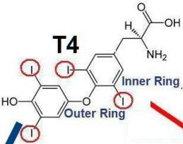
Thyroid Hormone (TH)
TH includes two related compounds: T4 (thyroxine) and T3 (triiodothyronine). TH increases metabolic rate, heat production, and is essential for normal development and function of many systems.
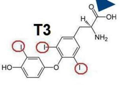
Synthesis, Transport, and Regulation of TH
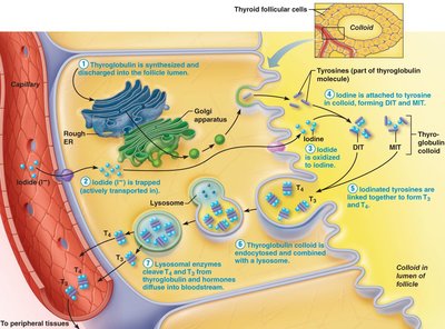
Thyroglobulin is synthesized and discharged into the follicle lumen. Iodide is actively transported, oxidized, and attached to tyrosine residues. Iodinated tyrosines combine to form T3 and T4, which are released into the bloodstream. T4 is converted to T3 in peripheral tissues. TH release is regulated by negative feedback involving TRH and TSH.
Homeostatic Imbalances of TH
Hyposecretion in adults: Myxedema; endemic goiter if due to iodine deficiency.
Hyposecretion in infants: Congenital Iodine Deficiency Syndrome (mental retardation, short stature).
Hypersecretion: Graves' disease (high metabolism, exophthalmos).
Calcitonin
Produced by parafollicular (C) cells, calcitonin lowers blood calcium by inhibiting osteoclast activity and stimulating calcium uptake into bone. Its physiological role in humans is minor.
Parathyroid Glands and Parathyroid Hormone (PTH)
Structure and Function
The parathyroid glands are small glands on the posterior thyroid. Chief cells secrete PTH, the most important hormone in calcium homeostasis.
PTH Actions
Stimulates osteoclasts to digest bone matrix, releasing Ca2+
Enhances reabsorption of Ca2+ and secretion of phosphate by kidneys
Promotes activation of vitamin D, increasing intestinal absorption of Ca2+
Homeostatic Imbalances of PTH
Hyperparathyroidism: Bone softening, kidney stones, nervous system depression.
Hypoparathyroidism: Tetany, respiratory paralysis, death.
Adrenal (Suprarenal) Glands
Structure and Layers
The adrenal glands are located atop the kidneys and consist of two regions: the adrenal cortex (glandular tissue) and adrenal medulla (nervous tissue).
Adrenal Cortex Hormones
Mineralocorticoids (e.g., aldosterone): Regulate Na+ and K+ balance, blood pressure, and volume.
Glucocorticoids (e.g., cortisol): Regulate metabolism, stress response, and immune function.
Gonadocorticoids: Sex hormones, mainly androgens.
Adrenal Medulla Hormones
Chromaffin cells secrete epinephrine (80%) and norepinephrine (20%), which mediate the fight-or-flight response by increasing blood glucose, heart rate, and blood pressure.
Pineal Gland
Melatonin Secretion
The pineal gland secretes melatonin, which regulates circadian rhythms, sexual maturation, and physiological cycles such as sleep and appetite.
Pancreas
Endocrine and Exocrine Functions
The pancreas contains both exocrine (acinar cells) and endocrine (islets of Langerhans) components. Alpha cells secrete glucagon (raises blood glucose), and beta cells secrete insulin (lowers blood glucose).
Glucagon and Insulin
Glucagon: Promotes glycogenolysis, gluconeogenesis, and glucose release from the liver.
Insulin: Enhances glucose uptake by cells, inhibits glycogenolysis and gluconeogenesis, and promotes fat and protein synthesis.
Homeostatic Imbalances of Insulin
Diabetes mellitus (DM): Due to insulin deficiency or inactivity; symptoms include polyuria, polydipsia, and polyphagia.
Hyperinsulinism: Excess insulin causes hypoglycemia, disorientation, and unconsciousness.
Gonads and Placenta
Ovaries and Testes
Ovaries: Produce estrogens and progesterone, regulating female reproductive development and cycles.
Testes: Produce testosterone, regulating male reproductive development and function.
Placenta: Secretes estrogens, progesterone, and human chorionic gonadotropin (hCG) during pregnancy.
Other Hormone-Producing Structures
Heart: Atrial natriuretic peptide (ANP) lowers blood pressure.
Gastrointestinal tract: Enteroendocrine cells secrete hormones regulating digestion.
Kidneys: Erythropoietin (EPO) stimulates red blood cell production; renin regulates blood pressure.
Skin: Produces cholecalciferol (vitamin D precursor).
Adipose tissue: Leptin regulates appetite and energy expenditure.
Skeleton: Osteocalcin enhances insulin secretion and sensitivity.
Thymus: Thymulin, thymopoietins, and thymosins regulate T lymphocyte development.
