 Back
BackThe Endocrine System: Structure, Function, and Clinical Significance
Study Guide - Smart Notes
 Tailored notes based on your materials, expanded with key definitions, examples, and context.
The Endocrine System
Overview
The endocrine system is a network of glands that secrete hormones directly into the bloodstream to regulate various physiological processes. These hormones control metabolism, growth, development, tissue function, sexual function, reproduction, sleep, and mood. 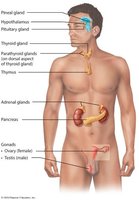
The Thyroid Gland
Location and Structure
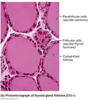
The thyroid gland is a butterfly-shaped organ located in the anterior neck, just inferior to the larynx and on the trachea. It consists of two lateral lobes connected by a median mass called the isthmus. The gland contains follicles, which are hollow spheres of epithelial follicular cells that produce the glycoprotein thyroglobulin. 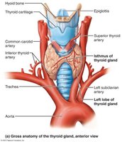
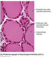
Colloid and Parafollicular Cells
The colloid is the fluid within the follicle lumen, containing thyroglobulin and iodine, serving as the precursor to thyroid hormone. Parafollicular cells, located outside the follicles, produce the hormone calcitonin.
Thyroid Hormone (TH)
Forms and Synthesis
Thyroid hormone is the body's major metabolic hormone and exists in two forms:
T4 (thyroxine): Consists of two tyrosine molecules with four bound iodine atoms; the major form secreted by the thyroid.
T3 (triiodothyronine): Consists of two tyrosines with three bound iodine atoms; more active at the tissue level.
Both are iodine-containing amine hormones. Synthesis involves several steps, including thyroglobulin production, iodide trapping, oxidation to iodine, attachment to tyrosine, and formation of T3 and T4. 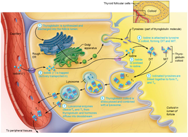
Transport and Regulation
T4 and T3 are transported by thyroxine-binding globulins (TBGs). T3 is ten times more active than T4. Peripheral tissues convert T4 to T3 by removing one iodine atom. Release of TH is regulated by negative feedback: falling TH levels stimulate TSH release, while rising TH levels inhibit TSH. 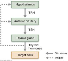
Major Effects of Thyroid Hormone
Increases basal metabolic rate and heat production (calorigenic effect)
Regulates tissue growth and development, especially skeletal and nervous systems
Maintains blood pressure by increasing adrenergic receptors in blood vessels
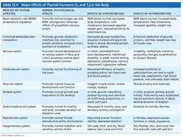
Process or System | Normal Effects | Effects of Hyposecretion | Effects of Hypersecretion |
|---|---|---|---|
Metabolic rate | Promotes normal BMR | BMR below normal, cold intolerance | BMR above normal, heat intolerance |
Carbohydrate/lipid/protein metabolism | Promotes glucose utilization | Impaired glucose metabolism | Enhanced catabolism of glucose |
Cardiovascular system | Promotes normal heart function | Decreased heart function | Increased heart function |
Nervous system | Promotes normal development | Impaired development | Hyperexcitability |
Muscular system | Promotes normal muscular development | Muscle weakness | Muscle tremor |
Gastrointestinal system | Promotes normal motility | Decreased motility | Increased motility |
Reproductive system | Promotes normal reproductive function | Impaired function | Impaired function |
Integumentary system | Promotes normal hydration | Dry, thick skin | Flushed, moist skin |
Clinical Homeostatic Imbalances of Thyroid Hormone
Hyposecretion
In adults, can lead to myxedema: low metabolic rate, thick/dry skin, puffy eyes, feeling chilled, constipation, edema, mental sluggishness, lethargy.
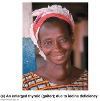
If due to iodine deficiency, a goiter may develop: thyroid enlarges as it attempts to produce more hormone.
Congenital Hypothyroidism
Usually caused by poor development of the thyroid gland in early childhood.
May present with weak cry, poor feeding, constipation, or prolonged jaundice.
TH replacement is crucial and lifelong.
Hypersecretion
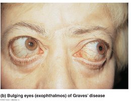
Most common type is Graves’ disease: autoimmune disorder where antibodies mimic TSH, stimulating TH release.
Symptoms: elevated metabolic rate, sweating, rapid and irregular heartbeats, nervousness, weight loss, exophthalmos (bulging eyes).
Treatment: surgical removal or radioactive iodine.
Calcitonin
Produced by parafollicular (C) cells in response to high Ca2+ levels.
Antagonist to parathyroid hormone (PTH).
At high doses, inhibits osteoclast activity and stimulates Ca2+ uptake into bone matrix.
The Parathyroid Glands
Location and Structure
Four to eight tiny yellow-brown glands embedded in the posterior aspect of the thyroid. Contain oxyphil cells (function unclear) and parathyroid cells that secrete parathyroid hormone (PTH), the most important hormone in Ca2+ homeostasis. 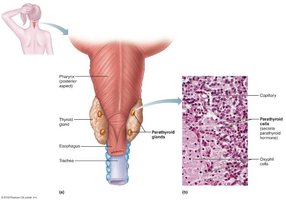
Functions of Parathyroid Hormone (PTH)
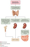
Stimulates osteoclasts to digest bone matrix and release Ca2+ to blood.
Enhances reabsorption of Ca2+ and secretion of phosphate by kidneys.
Promotes activation of vitamin D by kidneys, increasing absorption of Ca2+ by intestinal mucosa.
Clinical Homeostatic Imbalances of Parathyroid Hormone
Hyperparathyroidism
Usually due to parathyroid gland tumor.
Calcium leaches from bones, causing them to soften and deform.
Elevated Ca2+ depresses nervous system and contributes to kidney stones.
Osteitis fibrosa cystica: severe form resulting in easily fractured bones.
Hypoparathyroidism
Following gland trauma or removal can cause hypocalcemia.
Results in tetany, respiratory paralysis, and death.
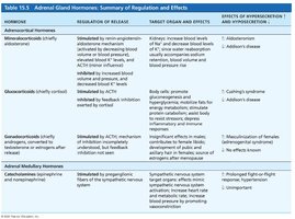
The Adrenal Glands
Location and Structure
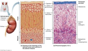
Paired, pyramid-shaped organs atop the kidneys, also called suprarenal glands. Structurally and functionally two glands in one:
Adrenal cortex: Three layers of glandular tissue synthesizing and secreting corticosteroids.
Adrenal medulla: Nervous tissue, part of the sympathetic nervous system.
Adrenal Cortex
Mineralocorticoids
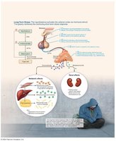
Regulate electrolyte concentrations (primarily Na+ and K+) in extracellular fluid.
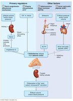
Aldosterone: Most potent mineralocorticoid; stimulates Na+ reabsorption and K+ elimination by kidneys.
Regulated by renin-angiotensin-aldosterone mechanism, plasma K+ concentration, ACTH, and atrial natriuretic peptide (ANP).
Clinical Imbalance: Aldosteronism
Hypersecretion usually due to adrenal tumors.
Results in hypertension, edema, and excretion of K+, leading to abnormal neurons and muscle function.
Glucocorticoids
Influence metabolism of most cells and help resist stressors.
Keep blood glucose levels constant and maintain blood pressure.
Cortisol: Only glucocorticoid in significant amounts in humans; released in response to ACTH.
Prime metabolic effect: gluconeogenesis (formation of glucose from fats and proteins).
Excessive levels depress cartilage/bone formation, inhibit inflammation, depress immune system, and disrupt normal functions.
Clinical Imbalances
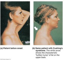
Cushing’s syndrome: Hypersecretion; causes "moon" face, "buffalo hump," immune suppression, and other disruptions.
Addison’s disease: Hyposecretion; deficits in glucocorticoids and mineralocorticoids, weight loss, dehydration, hypotension, and skin bronzing.
Gonadocorticoids
Weak androgens converted to testosterone or estrogens.
Contribute to puberty, sex drive in women, and source of estrogens in postmenopausal women.
Hypersecretion leads to adrenogenital syndrome (masculinization).
Adrenal Medulla
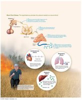
Medullary chromaffin cells synthesize catecholamines: epinephrine (80%) and norepinephrine (20%).
Effects: vasoconstriction, increased heart rate, increased blood glucose, blood diverted to brain, heart, and skeletal muscle.
Epinephrine stimulates metabolic activities; norepinephrine influences peripheral vasoconstriction and blood pressure.
Responses to stressors are brief.
Clinical Imbalance
Hypersecretion leads to symptoms of uncontrolled sympathetic nervous system (hyperglycemia, rapid heartbeat, hypertension, nervousness, sweating).
Can be due to pheochromocytoma (tumor of medullary chromaffin cells).
Hormone | Regulation of Release | Target Organ and Effects | Effects of Hyper/Hyposecretion |
|---|---|---|---|
Mineralocorticoids (aldosterone) | Stimulated by renin-angiotensin mechanism, increased K+, ACTH, inhibited by ANP | Kidney: increases blood Na+, decreases blood K+, increases blood volume and pressure | Hyper: Aldosteronism; Hypo: Addison's disease |
Glucocorticoids (cortisol) | Stimulated by ACTH, inhibited by feedback | Body cells: promotes gluconeogenesis, resists stress, depresses immune system | Hyper: Cushing's syndrome; Hypo: Addison's disease |
Gonadocorticoids (androgens) | Stimulated by ACTH | Reproductive organs: onset of puberty, sex drive | Hyper: Masculinization; Hypo: No effects |
Adrenal medullary hormones (epinephrine, norepinephrine) | Stimulated by preganglionic sympathetic fibers | Heart, blood vessels, liver: fight-or-flight response | Hyper: Prolonged fight-or-flight; Hypo: No effects |
The Pineal Gland
Structure and Function
Small gland hanging from the roof of the third ventricle. Pinealocytes secrete melatonin, derived from serotonin. Melatonin affects timing of sexual maturation, day/night cycles, physiological rhythms, and production of antioxidant molecules.
The Pancreas
Location and Structure
Triangular gland located partially behind the stomach. Contains both exocrine (acinar cells) and endocrine (islets of Langerhans) cells. 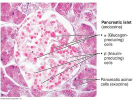
Hormones of the Pancreas
Alpha (α) cells: Produce glucagon (hyperglycemic hormone).
Beta (β) cells: Produce insulin (hypoglycemic hormone).
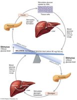
Glucagon
Triggered by decreased blood glucose, rising amino acids, or sympathetic stimulation.
Raises blood glucose by targeting the liver for glycogenolysis and gluconeogenesis.
Insulin
Secreted when blood glucose increases.
Lowers blood glucose by enhancing transport into fat and muscle cells, inhibiting glycogen breakdown, and inhibiting conversion of amino acids/fats to glucose.
Triggers cells to oxidize glucose for ATP, polymerize glucose to glycogen, and convert glucose to fat.
Clinical Homeostatic Imbalances of Pancreatic Hormones
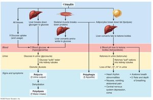
Diabetes Mellitus (DM)
Type 1: Hyposecretion of insulin
Type 2: Hypoactivity of insulin
Three cardinal signs: polyuria (excess urine), polydipsia (excess thirst), polyphagia (excess hunger)
Fats used as fuel, causing lipidemia and formation of ketones (ketoacidosis)
Untreated ketoacidosis can lead to coma and death
Hyperinsulinism
Excessive insulin secretion causes hypoglycemia (low blood glucose)
Symptoms: anxiety, nervousness, disorientation, unconsciousness, death
Treatment: sugar ingestion
The Gonads and Placenta
Ovaries
Produce estrogens and progesterone
Estrogen: maturation of reproductive organs, secondary sexual characteristics
Progesterone: breast development, cyclic changes in uterine mucosa
Testes
Produce testosterone
Initiates maturation of male reproductive organs, secondary sexual characteristics, sex drive
Necessary for normal sperm production
Placenta
Secretes estrogens, progesterone, and human chorionic gonadotropin (hCG)
Hormone Secretion by Other Organs
Adipose Tissue
Leptin: appetite control
Resistin: insulin antagonist
Adiponectin: enhances sensitivity to insulin
Gastrointestinal Tract
Gastrin: stimulates HCl release
Ghrelin: stimulates food intake
Secretin: stimulates liver and pancreas
Cholecystokinin (CCK): activates pancreas, gallbladder
Incretins: enhance insulin release, inhibit glucagon
Heart
Atrial natriuretic peptide (ANP): decreases blood Na+, blood pressure, and blood volume
Kidneys
Erythropoietin: signals production of red blood cells
Renin: initiates renin-angiotensin-aldosterone mechanism
Skeleton
Osteocalcin: prods pancreas to secrete more insulin, restricts fat storage, improves glucose handling
Skin
Cholecalciferol: precursor of vitamin D
Calcitriol: active form of vitamin D, helps absorb calcium, modulates immunity
Thymus
Thymulin, thymopoietins, thymosins: involved in T lymphocyte development
