 Back
BackThe Heart: Structure and Function – Anatomy & Physiology Study Notes
Study Guide - Smart Notes
 Tailored notes based on your materials, expanded with key definitions, examples, and context.
The Heart
Introduction to the Cardiovascular System
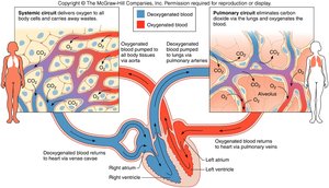
The cardiovascular system is responsible for transporting blood, nutrients, gases, and wastes throughout the body. The heart acts as the central pump, maintaining circulation through two main circuits: the systemic and pulmonary circuits.
Systemic circuit: Delivers oxygenated blood to body tissues and returns deoxygenated blood to the heart.
Pulmonary circuit: Carries deoxygenated blood to the lungs for gas exchange and returns oxygenated blood to the heart.
Section 1: Structures of the Heart
Location of the Heart
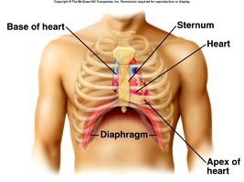
The heart is located in the thoracic cavity, posterior to the sternum, medial to the lungs, and anterior to the vertebral column. Its base lies beneath the second rib, and the apex rests at the fifth intercostal space on the diaphragm.
Base: Superior aspect, beneath the second rib.
Apex: Inferior, pointed end at the fifth intercostal space.
Coverings of the Heart (Pericardium)
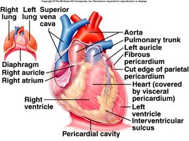
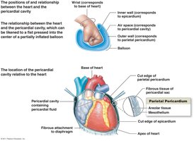
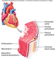
The heart is enclosed by the pericardium, a double-walled sac that protects and anchors the heart while reducing friction during heartbeats.
Fibrous pericardium: Outermost, tough connective tissue layer.
Parietal pericardium: Lines the inner surface of the fibrous pericardium.
Visceral pericardium (Epicardium): Covers the heart surface.
Pericardial cavity: Space between parietal and visceral layers, filled with serous fluid to reduce friction.
Wall of the Heart
The heart wall consists of three layers, each with distinct functions:
Epicardium (Visceral pericardium): Outer layer, reduces friction.
Myocardium: Thick, muscular middle layer responsible for contraction and pumping blood.
Endocardium: Inner epithelial and connective tissue layer, forms a protective lining inside the chambers.
Heart Chambers
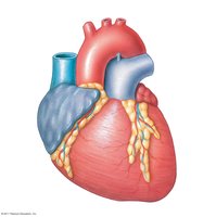
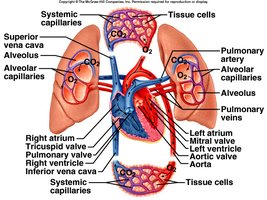
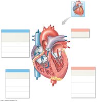
The heart contains four chambers: two atria (upper chambers) and two ventricles (lower chambers). The right side receives deoxygenated blood and pumps it to the lungs, while the left side receives oxygenated blood and pumps it to the body.
Right atrium: Receives blood from the superior and inferior vena cava and coronary sinus.
Right ventricle: Pumps blood to the lungs via the pulmonary arteries.
Left atrium: Receives blood from the pulmonary veins.
Left ventricle: Pumps blood to the systemic circuit via the aorta.
Heart Valves
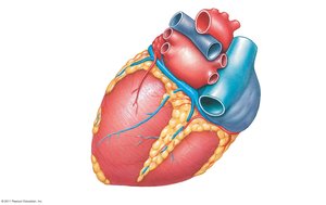
Heart valves ensure unidirectional blood flow through the heart and prevent backflow. There are two types: atrioventricular (AV) valves and semilunar valves.
Atrioventricular (AV) valves: Tricuspid (right) and mitral/bicuspid (left) valves, located between atria and ventricles.
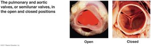
Semilunar valves: Pulmonary (right) and aortic (left) valves, located at the exits of the ventricles.
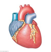
Structure and Function of Heart Valves
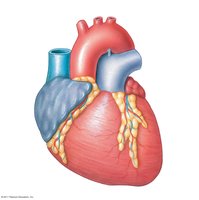
Tricuspid valve: Three cusps, prevents backflow into right atrium.
Mitral (bicuspid) valve: Two cusps, prevents backflow into left atrium.
Pulmonary valve: Three cusps, prevents backflow into right ventricle.
Aortic valve: Three cusps, prevents backflow into left ventricle.
Chordae tendineae: Tendinous cords that anchor AV valve cusps to papillary muscles, preventing prolapse.
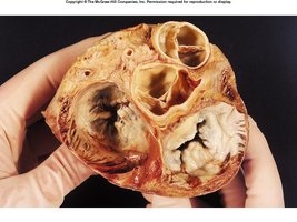
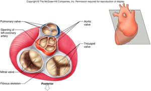
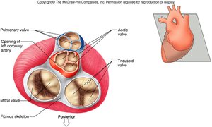
Valve | Location | Function |
|---|---|---|
Tricuspid valve | Right atrioventricular orifice | Prevents blood from moving from right ventricle into right atrium during ventricular contraction |
Pulmonary valve | Entrance to pulmonary trunk | Prevents blood from moving from pulmonary trunk into right ventricle during ventricular relaxation |
Mitral valve | Left atrioventricular orifice | Prevents blood from moving from left ventricle into left atrium during ventricular contraction |
Aortic valve | Entrance to aorta | Prevents blood from moving from aorta into left ventricle during ventricular relaxation |
Path of Blood Through the Heart
Blood flows through the heart in a specific sequence, ensuring oxygenation and systemic delivery:
Blood from systemic circuit enters right atrium via venae cavae and coronary sinus.
Passes through tricuspid valve into right ventricle.
Pumped through pulmonary valve into pulmonary arteries to the lungs.
Oxygenated blood returns via pulmonary veins to left atrium.
Passes through mitral valve into left ventricle.
Pumped through aortic valve into aorta and systemic circuit.
Blood Supply to the Heart (Coronary Circulation)
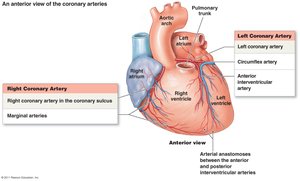
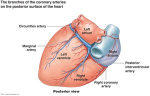
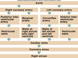
The heart muscle (myocardium) receives its own blood supply via the coronary arteries, which branch from the aorta. Venous blood is collected by cardiac veins and returned to the right atrium via the coronary sinus.
Right coronary artery: Branches into posterior interventricular and marginal arteries.
Left coronary artery: Branches into anterior interventricular and circumflex arteries.
Coronary sinus: Main vein returning deoxygenated blood from myocardium to right atrium.
Section 2: Physiology of the Heart
Cardiac Conduction System
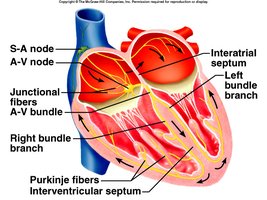
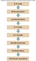
The cardiac conduction system coordinates the heartbeat, ensuring efficient pumping. Specialized cardiac muscle cells generate and transmit electrical impulses.
Sinoatrial (SA) node: Pacemaker, initiates heartbeat.
Atrioventricular (AV) node: Delays impulse, allowing atria to contract before ventricles.
AV bundle (Bundle of His): Conducts impulse to ventricles.
Bundle branches and Purkinje fibers: Distribute impulse through ventricles, causing contraction.
Electrocardiogram (ECG)
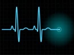
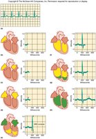
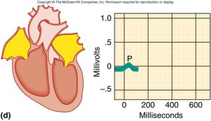
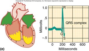
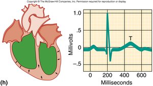
An ECG records the electrical changes in the myocardium during the cardiac cycle. It is used to assess heart rhythm and detect abnormalities.
P wave: Atrial depolarization (contraction).
QRS complex: Ventricular depolarization (contraction).
T wave: Ventricular repolarization (relaxation).
Heart Actions: Systole and Diastole
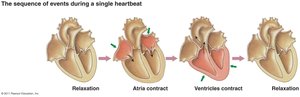
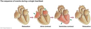
The cardiac cycle consists of alternating periods of contraction (systole) and relaxation (diastole) in the atria and ventricles.
Atrial systole: Atria contract, ventricles relax, AV valves open, semilunar valves closed.
Ventricular systole: Ventricles contract, atria relax, AV valves closed, semilunar valves open.
Cardiac Muscle Fibers
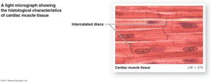
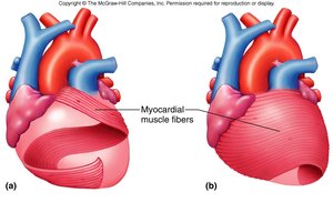
Cardiac muscle fibers are interconnected by intercalated discs, allowing the heart to function as a syncytium (a coordinated unit). The arrangement of fibers in the ventricular walls enables efficient, twisting contractions.
Atrial syncytium: Mass of merging cells in the atria.
Ventricular syncytium: Mass of merging cells in the ventricles.
Heart Sounds
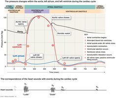
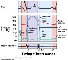
Heart sounds are produced by the closing of heart valves during the cardiac cycle. They are best heard at specific auscultation points on the chest.
Lubb (S1): First heart sound, caused by closure of AV valves during ventricular systole.
Dubb (S2): Second heart sound, caused by closure of semilunar valves during ventricular diastole.
Additional sounds (S3, S4): May be present in certain conditions.
Heart murmur: Abnormal sound due to leaking valve.

Cardiac Output and Blood Pressure
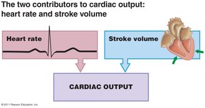
Cardiac output is the volume of blood pumped by each ventricle per minute. It is a key determinant of blood pressure, which is the force exerted by blood on vessel walls.
Cardiac Output (CO):
Stroke Volume (SV):
Blood Pressure (BP):
Where: HR = Heart Rate, EDV = End Diastolic Volume, ESV = End Systolic Volume, PR = Peripheral Resistance
Combined equation:
Additional info: Regulation of cardiac output and blood pressure involves neural, hormonal, and intrinsic mechanisms to maintain homeostasis.
