 Back
BackThe Heart: Structure, Function, and Disease
Study Guide - Smart Notes
 Tailored notes based on your materials, expanded with key definitions, examples, and context.
The Heart: Structure, Function, and Disease
Location, Size, and Position of the Heart
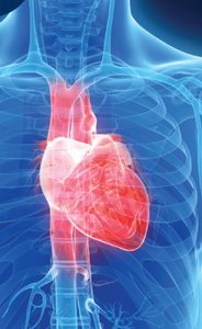
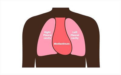
The heart is a muscular organ located in the mediastinum, the central compartment of the thoracic cavity. Approximately two-thirds of the heart's mass lies to the left of the body's midline, with the apex resting on the diaphragm. Its size is comparable to a closed fist, and its shape is roughly triangular.
Position: The heart lies between the sternum (anteriorly) and the thoracic vertebrae (posteriorly).
Clinical Relevance: Rhythmic compression of the heart between the sternum and vertebrae can maintain blood flow during cardiac arrest (cardiopulmonary resuscitation, CPR).
Apical Heartbeat: The apex is the best location to listen for heart sounds, typically between the fifth and sixth ribs at the midclavicular line.
Functional Anatomy of the Heart
The heart consists of four chambers: two atria (upper, receiving chambers) and two ventricles (lower, discharging chambers). The walls of these chambers are composed of cardiac muscle tissue called the myocardium. The inner lining is the endocardium, which provides a smooth surface for blood flow and helps prevent clot formation.
Atria: Right and left atria receive blood returning to the heart.
Ventricles: Right and left ventricles pump blood out of the heart.
Endocarditis: Inflammation of the endocardium, which can lead to thrombus (clot) formation.
Pericardium and Pericarditis
The pericardium is a two-layered fibrous sac that surrounds the heart, consisting of the inner visceral pericardium (epicardium) and the outer parietal pericardium. Between these layers is a lubricated space containing pericardial fluid, which reduces friction during heartbeats. Inflammation of the pericardium is called pericarditis, and fluid accumulation can lead to cardiac tamponade, a life-threatening compression of the heart.
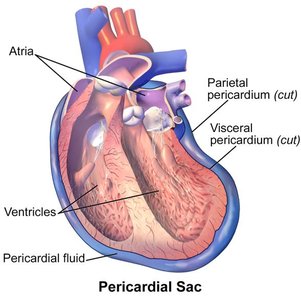
Pericardial Fluid: Prevents friction between the two pericardial layers during heartbeats.
Internal Structure of the Heart
The heart's internal anatomy includes the four chambers, valves, and associated structures. The myocardium forms the bulk of the heart wall, while the endocardium lines the chambers. The pericardium surrounds the heart, providing protection and reducing friction.
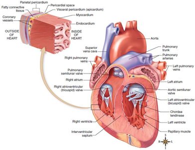
Valves: Ensure unidirectional blood flow through the heart.
Septum: Divides the right and left sides of the heart.
Heart Action and Valves
Cardiac Cycle: Systole and Diastole
The cardiac cycle consists of alternating periods of contraction (systole) and relaxation (diastole). During atrial systole, the atria contract to fill the ventricles. Ventricular systole follows, pumping blood out of the heart. Valves control the direction of blood flow and prevent backflow.
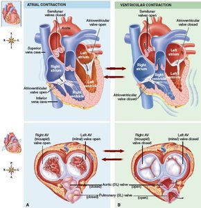
Systole: Contraction phase of the heart.
Diastole: Relaxation phase of the heart.
Valves: Atrioventricular (AV) valves (tricuspid and bicuspid/mitral) and semilunar (SL) valves (pulmonary and aortic).
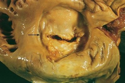
Heart Valve Disorders
Valves maintain one-way blood flow. Disorders include:
Incompetent Valves: Allow backflow of blood (regurgitation).
Stenosed Valves: Narrowed, restricting blood flow.
Rheumatic Heart Disease: Cardiac damage from a delayed inflammatory response to streptococcal infection.
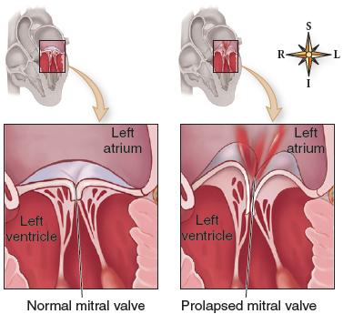
Mitral Valve Prolapse (MVP): The mitral valve bulges into the left atrium during ventricular contraction, causing incompetence.
Heart Sounds
Each heartbeat produces two main sounds:
First sound (lub): Closure of AV valves during ventricular contraction.
Second sound (dup): Closure of SL valves during ventricular relaxation.
Heart Murmurs: Abnormal sounds, often due to valve disorders.
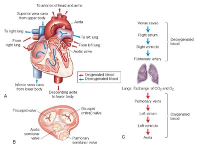
Blood Flow Through the Heart and Cardiovascular System
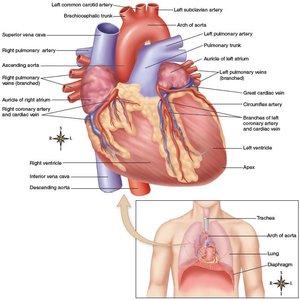
Pathway of Blood Through the Heart
The heart acts as two separate pumps: the right side pumps blood to the lungs (pulmonary circulation), and the left side pumps blood to the body (systemic circulation).
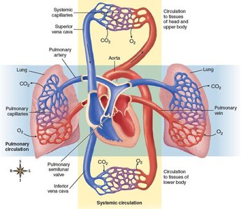
Venous blood enters the right atrium via the superior and inferior venae cavae.
Blood passes through the tricuspid valve to the right ventricle.
From the right ventricle, blood is pumped through the pulmonary semilunar valve to the pulmonary artery and lungs.
Oxygenated blood returns from the lungs to the left atrium, passes through the bicuspid (mitral) valve to the left ventricle.
The left ventricle pumps blood through the aortic semilunar valve into the aorta for systemic distribution.
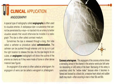
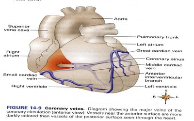
Blood Supply to the Heart Muscle (Coronary Circulation)
The myocardium receives oxygen and nutrients from the right and left coronary arteries. Blockage of these arteries can cause myocardial infarction (heart attack).
Coronary Thrombosis/Embolism: Blood clot blocking a coronary artery, leading to tissue death (myocardial infarction).
Atherosclerosis: Lipid buildup in arteries, restricting blood flow.
Angina Pectoris: Chest pain from inadequate oxygen supply to the myocardium.
Coronary Bypass and Angioplasty
Coronary bypass surgery uses vessels from other parts of the body to bypass blocked coronary arteries. Angioplasty is a less invasive procedure to open blocked arteries using a device.
Cardiac Cycle and Output
Cardiac Cycle
Each heartbeat is a cardiac cycle, averaging 72 beats per minute. Each cycle lasts about 0.8 seconds and includes systole and diastole.
Stroke Volume (SV): Volume of blood ejected from one ventricle per beat.
Cardiac Output (CO): Amount of blood pumped by one ventricle per minute.
Equation for cardiac output:
HR: Heart rate (beats per minute)
SV: Stroke volume (volume per beat)
Conduction System of the Heart and Electrocardiography
Conduction System
The heart's conduction system coordinates contraction:
SA (Sinoatrial) Node: Pacemaker, initiates impulse.
AV (Atrioventricular) Node: Receives impulse from SA node.
AV Bundle (Bundle of His): Conducts impulse to ventricles.
Purkinje Fibers: Distribute impulse through ventricular myocardium.
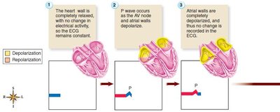
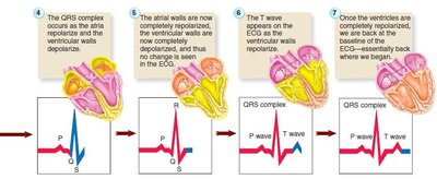
Electrocardiogram (ECG/EKG)
An ECG records the electrical activity of the heart. The normal ECG has three main deflections:
P wave: Atrial depolarization
QRS complex: Ventricular depolarization
T wave: Ventricular repolarization
Cardiac Dysrhythmias
Types of Dysrhythmias
Cardiac dysrhythmias are abnormalities in heart rhythm:
Bradycardia: Slow heart rate (<60 bpm)
Tachycardia: Rapid heart rate (>100 bpm)
Sinus Dysrhythmia: Variation in heart rate during breathing
Premature Contraction (Extrasystole): Early contraction
Fibrillation: Uncoordinated contraction, no effective pumping
Fibrillation can be life-threatening, especially ventricular fibrillation. Defibrillation (electric shock) can restore normal rhythm. Atrial ablation is a procedure to treat atrial fibrillation by destroying abnormal conduction pathways.
Heart Failure
Types and Causes
Heart failure is the inability of the heart to pump enough blood to sustain life. It can result from various heart diseases, including cardiomyopathy, valve disorders, and myocardial infarction.
Right Heart Failure: Often due to left heart failure; leads to systemic congestion.
Left Heart Failure (Congestive Heart Failure, CHF): Inability of the left ventricle to pump effectively, causing pulmonary and systemic congestion.
Treatment: Heart transplants and artificial hearts are options for end-stage heart failure.
Review Questions
Question | Answer |
|---|---|
What is the thin, smooth lining of each heart chamber called? | Endocardium |
What is cardiac damage from a delayed inflammatory response to streptococcal infection called? | Rheumatic heart disease |
What often causes abnormal heart sounds (murmurs)? | Valve disorders |
Where does impulse conduction in the heart normally start? | Sinoatrial (SA) node |
What do the deflections of an ECG tracing represent? | Electrical activity regulating atrial and ventricular contraction and relaxation |
What is sinus dysrhythmia? | Variation in heart rate during the breathing cycle |
What can affect cardiac stroke volume? | Hormones, ion imbalances, valve disorders (all of these) |
