Back
The Immune System: Innate and Adaptive Body Defenses
Study Guide - Smart Notes
 Tailored notes based on your materials, expanded with key definitions, examples, and context.
Innate (Nonspecific) Defenses
Species Specificity
Some organisms are naturally resistant to diseases that affect other species. This resistance is due to unique physiological or biochemical characteristics that prevent infection or disease progression.
Key Point: Species-specific barriers are the first line of defense, preventing cross-species infections.
Example: Humans are not susceptible to certain animal diseases, and vice versa.
Mechanical Barriers (First Line of Defense)
The skin and mucous membranes act as physical barriers to pathogens, blocking their entry into the body.
Key Point: These barriers are always present and act against a wide range of assaults, including bacteria, viruses, burns, and cuts.
Example: Intact skin prevents most pathogens from entering the body.
Chemical Barriers (Second Line of Defense)
Enzymes and proteins in body fluids provide chemical protection against pathogens.
Interferons: Stimulate phagocytosis and interfere with viral replication.
Defensins: Disrupt microbial membranes, crippling microbes.
Complement: Enhances inflammation, attracts phagocytes, and promotes phagocytosis.
Collectins: Provide broad protection against bacteria, yeast, and some viruses.
Transferrin: Inhibits bacterial growth by reducing available iron (Fe).
Natural Killer (NK) Cells (Second Line of Defense)
NK cells are a type of lymphocyte that target virus-infected and cancerous cells. They secrete cytolytic substances called perforins, which destroy infected cells.
Key Point: NK cells act similarly to defensins by rupturing cell membranes.
Inflammation (Second Line of Defense)
Inflammation is a localized response to injury or infection, characterized by redness, swelling, heat, and pain.
Rubor: Redness
Tumor: Swelling
Dolor: Pain
Calor: Heat
Phagocytosis (Second Line of Defense)
Phagocytic cells, such as neutrophils and monocytes, ingest and destroy pathogens. Chemotaxis is the process by which these cells are attracted to the site of injury or infection.
Key Point: Phagocytosis is essential for clearing pathogens and cellular debris.
Fever (Second Line of Defense)
A fever is triggered by the release of interleukin-1 (IL-1) from lymphocytes in response to infection. Elevated body temperature can inhibit pathogen growth and enhance immune responses.
Key Point: Fever is a systemic response that supports the immune system.
Adaptive (Specific) Defenses or Immunity (Third Line of Defense)
Overview of Adaptive Immunity
Adaptive immunity is the body's third line of defense, characterized by specificity and memory. It distinguishes between self and non-self molecules, primarily through the recognition of antigens.
Antigen: A molecule on the surface of cells that can elicit an immune response by stimulating antibody production. The most effective antigens are large and complex.
Lymphocyte Origins
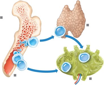
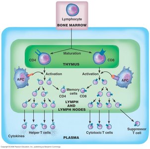
Lymphocytes originate from stem cells in the red bone marrow. They differentiate into two main types: B cells and T cells. B cells mature in the bone marrow, while T cells mature in the thymus. Both cell types circulate through the blood and lymphatic organs.
T Cells and the Cellular Immune Response
T cells are central to the cellular immune response. Activation requires two signals: recognition of an antigen presented by an antigen-presenting cell (APC) with MHC molecules, and a costimulatory signal (often cytokines like interleukin-2).
Helper T cells (CD4): Activate other immune cells, including cytotoxic T cells and B cells. Essential for initiating immune responses. Targeted by HIV.
Cytotoxic T cells (CD8): Destroy infected or abnormal cells by releasing granules that cause cell lysis. Require signals from helper T cells.
Memory T cells: Persist after an infection to provide a rapid response upon re-exposure to the same antigen.
B Cells and the Humoral Immune Response
B cells are activated when their receptors bind to specific antigens, often with help from helper T cells. Activated B cells differentiate into plasma cells, which secrete antibodies (immunoglobulins), or become memory B cells for future responses.
Key Point: Antibodies are proteins that specifically bind to antigens, marking them for destruction.
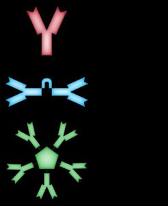
Major Immunoglobulins (Antibody Classes)
IgG: Crosses the placenta, provides passive immunity, effective against bacteria, viruses, and toxins.
IgM: First antibody produced in response to infection; includes anti-A and anti-B antibodies.
IgA: Found in body fluids such as saliva, tears, and breast milk.
IgD: Present on the surface of B cells; involved in B cell activation.
IgE: Involved in allergic reactions (e.g., hay fever).
How Antibodies Work
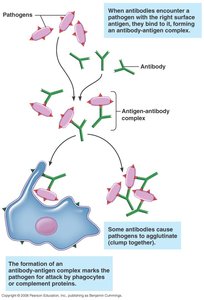
Antibodies bind to antigens, forming antigen-antibody complexes. This process can neutralize pathogens, cause them to clump (agglutinate), or mark them for destruction by phagocytes or complement proteins.
Antibody Structure
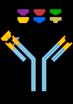
Antibodies have a Y-shaped structure with variable regions that bind specific antigens. The constant region determines the antibody class and mediates immune functions.
Primary and Secondary Immune Responses
The primary immune response occurs after the first exposure to an antigen and is slower and less robust. The secondary response, upon re-exposure, is faster and stronger due to memory cells.
Key Point: Immunological memory is the basis for effective vaccination.
Practical Classification of Immunity
Naturally Acquired Active Immunity: Results from exposure to a live pathogen.
Artificially Acquired Active Immunity: Results from vaccination with weakened or dead pathogens.
Artificially Acquired Passive Immunity: Involves injection of antibodies.
Naturally Acquired Passive Immunity: Antibodies are transferred from mother to child via placenta or breast milk.
Hypersensitivity (Allergic) Reactions
Type I: Immediate reaction (minutes); symptoms include hives, hay fever, asthma, anaphylactic shock.
Type II: Cytotoxic reaction (1-3 hours); e.g., transfusion reactions.
Type III: Immune-complex reaction (1-3 hours); antibody complexes cause tissue damage (e.g., rheumatoid arthritis).
Type IV: Delayed reaction (about 48 hours); e.g., contact dermatitis from poison ivy.
Transplantation and Tissue Rejection
Tissue rejection occurs when the recipient's immune system recognizes donor tissue as foreign. Graft-versus-host disease (GVHD) can occur if donor immune cells attack the recipient. Matching MHC antigens and using immunosuppressive drugs help prevent rejection.
Key Point: Immune suppression increases the risk of infection but is necessary to prevent rejection.
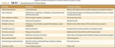
Autoimmunity
Autoimmune diseases occur when the immune system fails to distinguish self from non-self, producing autoantibodies and cytotoxic T cells that attack the body's own tissues.
Disorder | Symptoms | Antibodies Against |
|---|---|---|
Glomerulonephritis | Lower back pain | Kidney cell antigens that resemble streptococcal bacteria antigens |
Graves disease | Restlessness, weight loss, irritability, increased heart rate and blood pressure | Thyroid gland antigens near thyroid-stimulating hormone receptor; causing overactivity |
Type 1 diabetes mellitus | Thirst, hunger, weakness, emaciation | Pancreatic beta cells |
Hemolytic anemia | Fatigue and weakness | Red blood cells |
Multiple sclerosis | Weakness, incoordination, speech disturbances, visual complaints | Myelin in the white matter of the central nervous system |
Myasthenia gravis | Muscle weakness | Receptors for acetylcholine on skeletal muscle |
Pernicious anemia | Fatigue and weakness | Binding site for vitamin B12 on cells lining stomach |
Rheumatic fever | Weakness, aches, shortness of breath | Heart valve cell antigens that resemble streptococcal bacteria antigens |
Rheumatoid arthritis | Joint pain and deformity | Cells lining joints |
Systemic lupus erythematosus | Red rash on face, prolonged fever, weakness, kidney damage, joint pain | Connective tissue |
Ulcerative colitis | Lower abdominal pain | Colon cells |