 Back
BackThe Immune System: Structure, Function, and Cellular Components
Study Guide - Smart Notes
 Tailored notes based on your materials, expanded with key definitions, examples, and context.
The Immune System
Role and Functions of the Immune System
The immune system is a complex network that protects the body from damage caused by pathogens, abnormal cells, and foreign substances. Its primary functions include defense against infectious agents, removal of dead or damaged cells, and recognition and elimination of abnormal self cells such as tumor cells.
Immunity: The body's ability to protect itself from disease and harmful agents. The term derives from Latin 'immunis' meaning exempt.
Self vs. Non-self: The immune system distinguishes between the body's own cells and foreign entities, including pathogens (bacteria, viruses, parasites, fungi), transplanted organs, transfused blood, environmental pollutants, and cancer cells.
Main Functions:
Scavenging and removing dead or damaged cells
Protecting the body from pathogens
Recognizing and removing abnormal own cells (e.g., tumor cells)
Pathogens and Non-infectious Substances
Pathogens are microorganisms capable of causing disease, while non-infectious substances can also elicit immune responses.
Microorganisms:
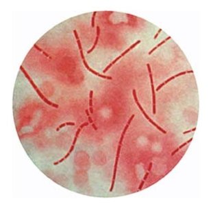
Bacteria (e.g., Staphylococcus, Salmonella, Helicobacter)
Viruses (e.g., Zika, COVID-19, Polio, HIV)
Parasites (e.g., malaria, tapeworms)
Non-infectious Substances:
Pollen
Chemicals (smoke, drugs)
Food
Historical Milestones in Microbiology
The study of microorganisms and their role in disease has evolved through key discoveries:
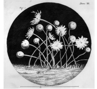
1665: Robert Hooke publishes the first depiction of a microorganism (microfungus Mucor).
1676: Antoni van Leeuwenhoek discovers bacteria in water.
1884: Robert Koch formulates the germ theory of disease, establishing criteria for linking microorganisms to specific diseases.
Immunogen, Antigen, and Epitope
Definitions and Relationships
Understanding the molecular basis of immune recognition is essential for immunology.
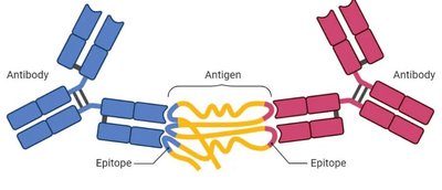
Immunogen: Any substance that triggers an immune response.
Antigen: An immunogen that binds with the products of the immune response (e.g., antibodies).
Epitope: The specific part of an antigen that binds to an antibody or antigen receptor on immune cells (B or T cells). Binding occurs only if the structures are complementary.
Anatomy of the Immune System
Lymphoid Tissues
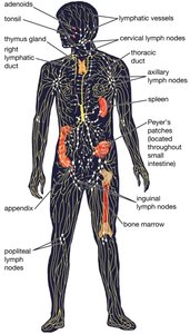
The immune system is composed of specialized tissues and organs where immune cells are generated, mature, and interact with pathogens.
Primary Lymphoid Tissues: Sites of immune cell generation and maturation.
Bone marrow
Thymus
Secondary Lymphoid Tissues: Sites of immune cell interaction with pathogens.
Encapsulated: Spleen, lymph nodes
Diffuse: Tonsils, Skin Associated Lymphoid Tissue (SALT), Mucosa Associated Lymphoid Tissue (MALT), Gut Associated Lymphoid Tissue (GALT)
Immune Cells (Leukocytes)
Immune cells, also known as leukocytes or white blood cells, are central to immune defense. They are produced in the bone marrow and circulate in blood and tissues.
White Blood Cell Production (Leukopoiesis):
Driven by colony-stimulating factors (CSFs), which are glycoproteins secreted by monocytes/macrophages, fibroblasts, and endothelial cells.
CSFs induce proliferation and differentiation of hematopoietic stem cells (HSCs) into specific WBC subtypes.
Hematopoietic Lineage and Immune Cell Types
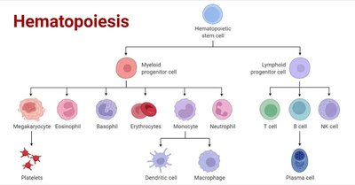
Hematopoiesis
Hematopoiesis is the process by which all blood cells are formed from pluripotent hematopoietic stem cells in the bone marrow. These stem cells can differentiate into multiple cell types, including immune cells.
Pluripotent Stem Cells: Uncommitted cells capable of self-renewal or differentiation.
Reduction of Differentiation Potential: As stem cells differentiate, their potential narrows to specific lineages.
Granulocytes
Granulocytes are a category of white blood cells characterized by granules in their cytoplasm. They play key roles in innate immunity and inflammation.
Types of Granulocytes:
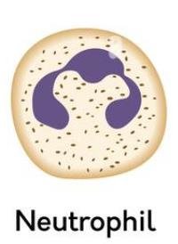
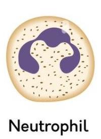
Neutrophils: Kill bacteria through phagocytosis; secrete pyrogens.
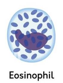
Eosinophils: Involved in allergic reactions and parasitic infections.
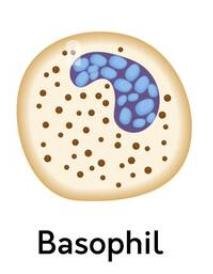
Basophils: Participate in innate immune response and inflammation; become mast cells in tissues.
Degranulation: When activated, granulocytes release their granule contents by exocytosis.

Granulocyte Staining and Functions
Granulocytes can be distinguished by their staining properties and functions:
Basophils: Stain with dark blue-basic hematoxylin dye; involved in inflammation.
Eosinophils: Stain with bright pink acidic dye eosin; involved in allergic reactions and parasitic infections.
Neutrophils: Do not stain strongly; kill bacteria through phagocytosis.
Monocytes and Macrophages
Monocytes are circulating white blood cells that extravasate into tissues and become macrophages. Macrophages patrol tissues for bacteria and debris, and are capable of killing up to 100 bacteria during their lifetime. They also function as antigen presenting cells (APCs).
Dendritic Cells
Dendritic cells are professional antigen presenting cells (APCs) that phagocytose, process, and display antigens for other immune cells. They link innate and adaptive immunity and are found primarily in barrier tissues such as skin.
Lymphocytes
B-cells, T-cells, and NK Cells
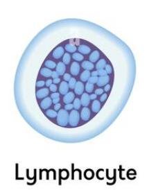
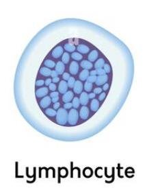
Lymphocytes are a major class of immune cells involved in adaptive immunity. They include B-cells, T-cells, and natural killer (NK) cells.
B-cells: Mature in the bone marrow; produce antibodies.
T-cells: Mature in the thymus; involved in cell-mediated immunity.
NK cells: Kill virus-infected and cancer cells without pre-stimulation.
Lymphocyte Development
Lymphocyte development involves selection for self-tolerance to prevent autoimmunity.
T-cell Development:
Progenitors originate in bone marrow and migrate to the thymus.
In the thymus, T-cells are trained to recognize specific antigens; self-reactive cells undergo apoptosis.
Only 2% of T-cell progenitors survive maturation.
Mature T-cells enter the bloodstream and lymphoid organs.
B-cell Development:
Progenitors originate and mature in bone marrow.
Immature B cells undergo selection for self-tolerance.
Naive B cells enter the bloodstream and further differentiate in peripheral lymphoid tissues.
Plasma cells develop from activated B cells and produce antibodies.
Antibodies and Immune Recognition
Antibody Structure and Classes
Antibodies are proteins produced by plasma cells (activated B cells) that bind to specific antigens. They consist of four polypeptide chains arranged in a Y shape, with antigen binding sites on the arms and a hinge region for flexibility.
Fc Region: Determines antibody class.
Classes of Immunoglobulins (Ig):
IgG: 75% of gamma globulins; maternal IgG provides passive immunity to infants.
IgA: Found in external secretions (saliva, breast milk, tears, mucus).
IgE: Involved in parasitic infections and allergies.
IgM: Early immune responses; activates complement.
IgD: Receptors on B cell surfaces; contribute to B cell activation.
Self-Recognition and Autoimmunity
Each B and T cell clone recognizes one particular antigen. Receptors are formed from millions of combinations. If receptors react with self-antigens, the clone is deleted during maturation, protecting against autoimmunity.
Autoimmune Diseases
Autoimmune diseases occur when self-tolerance fails and the body attacks itself. They are characterized by antibody production against specific epitopes, often have a genetic component, and may be triggered by infections.
~78% of autoimmune patients are female.
X-chromosome inactivating gene (Xist) may underlie female-biased autoimmunity.
Summary Table: Immune Cell Types and Functions
Cell Type | Location | Function |
|---|---|---|
Basophil | Blood, Tissues (as Mast cell) | Innate immune response, Inflammation |
Eosinophil | Blood, Tissues | Allergic reactions, Parasitic infections |
Neutrophil | Blood, Tissues (upon infection) | Kill bacteria through phagocytosis, Secrete pyrogens |
Monocyte | Blood | Patrol tissues, Become macrophages, Antigen presentation |
Lymphocyte | Blood, Lymphoid tissues | Adaptive immunity (B cells, T cells, NK cells) |
Conclusion
The immune system is a highly organized network of tissues, cells, and molecules that work together to defend the body against pathogens and maintain homeostasis. Understanding its structure and function is essential for comprehending human health and disease.
