 Back
BackThe Integumentary System: Structure, Function, and Clinical Aspects
Study Guide - Smart Notes
 Tailored notes based on your materials, expanded with key definitions, examples, and context.
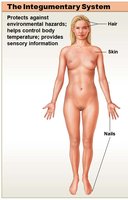
The Integumentary System
Overview and Major Components
The integumentary system is the body's largest organ system, serving as the primary barrier between the internal environment and the external world. It consists of the cutaneous membrane (skin) and accessory structures such as hair, nails, and exocrine glands.
Cutaneous membrane (skin): Composed of the epidermis and dermis.
Accessory structures: Include hair, nails, and exocrine glands (sebaceous and sweat glands).
General Functions of the Integument
Protection: Shields underlying tissues from mechanical, chemical, and biological damage; prevents fluid loss.
Temperature maintenance: Regulates heat exchange with the environment.
Synthesis and storage of nutrients: Epidermis synthesizes vitamin D3; dermis stores lipids.
Sensory reception: Detects touch, pressure, pain, and temperature.
Excretion and secretion: Glands excrete salts, water, and organic wastes; mammary glands secrete milk.
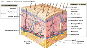
Structure of the Skin
Layers of the Skin
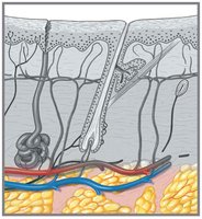
The skin consists of two main layers: the epidermis and the dermis. Beneath the dermis lies the subcutaneous layer (hypodermis), which is not technically part of the integument but is closely associated with it.
Epidermis: Superficial, avascular epithelial tissue.
Dermis: Deeper, vascular connective tissue layer.
Subcutaneous layer (hypodermis): Loose connective tissue and fat; stabilizes skin position.
The Epidermis
Characteristics and Cell Types
The epidermis is a stratified squamous epithelium, primarily composed of keratinocytes. It is avascular and relies on diffusion from the underlying dermis for nutrients and oxygen. The deepest cells are most metabolically active, while the outermost cells are dead and filled with keratin.
Keratinocytes: Main cell type, produce keratin for protection and water resistance.
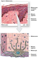
Melanocytes: Produce melanin pigment.
Merkel cells: Sensory receptors for touch.
Dendritic (Langerhans) cells: Immune defense.
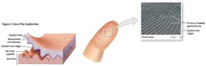
Thick Skin vs. Thin Skin
Thick skin: Found on palms and soles; has five layers (including stratum lucidum); thicker stratum corneum.
Thin skin: Covers most of the body; has four layers; thinner stratum corneum.
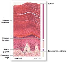
Strata of the Epidermis
The epidermis is organized into distinct layers (strata), each with specialized functions:
Stratum basale (germinativum): Deepest layer; contains stem cells, melanocytes, and Merkel cells; site of cell division.
Stratum spinosum: Several layers of keratinocytes; contains dendritic cells; cells connected by desmosomes.
Stratum granulosum: Cells stop dividing; begin producing keratin and keratohyalin granules.
Stratum lucidum: Present only in thick skin; clear, flattened, densely packed cells filled with keratin.
Stratum corneum: Outermost layer; 15–30 layers of dead, keratinized cells; provides barrier function.
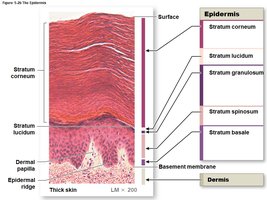
Epidermal Ridges and Dermal Papillae
Epidermal ridges (from the stratum basale) interlock with dermal papillae, increasing surface area for attachment and forming the basis for fingerprints.
Skin Color and Pigmentation
Carotene: Orange-yellow pigment from diet; accumulates in epidermal cells.
Melanin: Brown-black or red-yellow pigment produced by melanocytes; protects against UV radiation.
Dermal circulation: Oxygenated blood gives skin a pinkish hue; cyanosis (bluish color) indicates low oxygen.
Effects of Sunlight
Beneficial: Stimulates vitamin D3 synthesis, essential for calcium and phosphorus absorption.
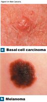
Detrimental: Excessive exposure increases risk of skin cancers (basal cell carcinoma, squamous cell carcinoma, malignant melanoma).
The Dermis
Structure and Layers
The dermis lies between the epidermis and hypodermis and provides structural strength and elasticity to the skin. It contains two layers:
Papillary layer: Superficial; areolar connective tissue; contains capillaries, lymphatics, and sensory neurons.
Reticular layer: Deeper; dense irregular connective tissue; contains collagen and elastic fibers, blood vessels, nerves, and accessory structures.
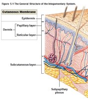
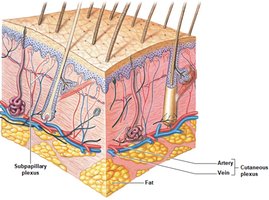
Blood Supply in the Dermis
Cutaneous plexus: Deep network of arteries and veins at the dermis-hypodermis boundary.
Subpapillary plexus: Supplies capillary loops at the epidermis-dermis boundary.
The Hypodermis (Subcutaneous Layer)
The hypodermis is a layer of areolar and adipose tissue beneath the dermis. It stabilizes the position of the skin, stores energy, and provides insulation. It is a common site for subcutaneous injections due to the absence of vital organs.
Accessory Structures of the Integument
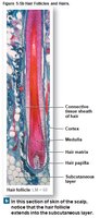
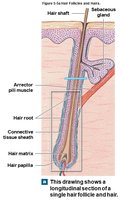
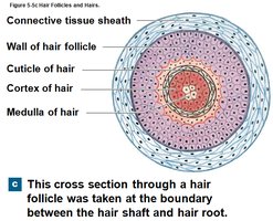
Hair and Hair Follicles
Hair is a nonliving, keratinized structure produced by hair follicles. It serves protective, sensory, and thermoregulatory functions.
Hair follicle: Extends into the dermis and hypodermis; contains all epidermal layers; base forms the hair matrix (site of cell division).
Hair structure: Root (below skin), shaft (above skin); composed of cuticle, cortex, and medulla.
Hair Growth and Color
Growth cycle: Active growth (2–5 years), followed by inactivity and shedding.
Color: Determined by type and amount of melanin; declines with age (gray/white hair).
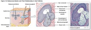
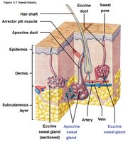
Exocrine Glands of the Skin
Sebaceous (oil) glands: Secrete sebum into hair follicles or directly onto skin; lubricates and protects.
Sudoriferous (sweat) glands: Two types:
Apocrine glands: Active at puberty; secrete into hair follicles in axillae, groin, and nipples; produce odorous secretion.
Eccrine (merocrine) glands: Widely distributed; secrete watery sweat directly onto skin; important for thermoregulation.
Modified glands: Mammary glands (milk), ceruminous glands (earwax).
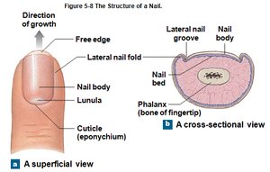
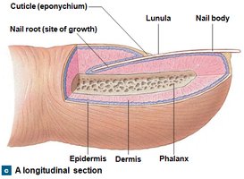
Nails
Nails are protective coverings on the dorsal surfaces of fingers and toes, composed of dense, keratinized cells. The nail root is the site of production, and the lunula is the pale crescent near the root.
Repair and Regeneration of the Integument
Phases of Skin Regeneration
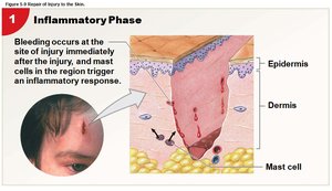
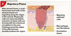
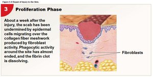
Skin repair involves four overlapping phases:
Inflammatory phase: Increased blood flow and phagocyte activity at the injury site.
Migratory phase: Formation of a scab; epithelial cells migrate to cover the wound; granulation tissue forms.
Proliferation phase: Fibroblasts produce collagen; the clot dissolves; tissue repair continues.
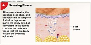
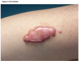
Scarring phase: Scar tissue forms; may result in keloids (thickened, shiny scars).
Burns and Their Effects
Classification by Depth
Burn Type | Depth | Features |
|---|---|---|
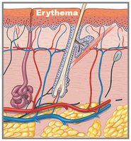
First-degree (partial-thickness) | Epidermis only | Erythema (redness), mild pain; e.g., sunburn |
Second-degree (partial-thickness) | Epidermis + part of dermis | Blistering, pain, swelling, possible scarring |
Third-degree (full-thickness) | Epidermis + dermis destroyed, may extend to hypodermis | Sensory nerves destroyed, requires grafting |
Effects of Burns
Fluid and electrolyte imbalance
Impaired thermoregulation
Increased risk of infection
Effects of Aging on the Integument
Thinner epidermis; increased risk of injury and infection
Reduced immune sensitivity
Decreased vitamin D3 production; weaker bones and muscles
Lower melanin production; increased sun sensitivity
Reduced glandular activity; drier, scaly skin
Thinning and graying of hair
Loss of skin elasticity; sagging and wrinkling
Slower skin repair
