Back
BackThe Integumentary System: Structure, Function, and Components
Study Guide - Smart Notes
 Tailored notes based on your materials, expanded with key definitions, examples, and context.
Chapter 4: The Integumentary System (Skin)
Overview and Functions
The integumentary system is the body's largest organ system, primarily composed of the skin and its accessory structures. It serves as the first line of defense and performs several vital physiological roles.
Physical protection: Shields internal tissues from mechanical damage, pathogens, and dehydration.
Regulation of body temperature: Maintains thermal homeostasis via sweat production and blood flow regulation.
Excretion: Removes metabolic wastes through sweat glands.
Nutrition: Synthesizes vitamin D3 in the epidermis upon UV exposure.
Sensation: Contains sensory receptors for touch, pressure, pain, and temperature.
Immune defense: Coordinates immune responses to pathogens and injury.
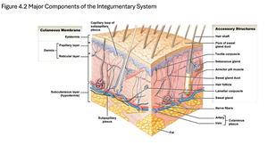
Major Components of the Skin
Cutaneous Membrane
The cutaneous membrane consists of two main layers: the epidermis and the dermis.
Epidermis: Outermost layer, composed of keratinized stratified squamous epithelium. Functions include protection, water retention, and vitamin D3 synthesis.
Dermis: Lies beneath the epidermis and is divided into two layers:
Papillary layer: Contains capillaries, nerves, and loose connective tissue; nourishes and supports the epidermis.
Reticular layer: Dense connective tissue with blood vessels, hair follicles, and glands; provides strength, elasticity, and houses sensory receptors.
Accessory Structures
Accessory structures originate in the dermis and extend through the epidermis to the skin surface.
Hair follicles: Produce hair for protection and sensory input.
Nails: Protect and support the tips of fingers and toes.
Glands: Include sebaceous (oil) and sweat glands, which assist in thermoregulation, excretion, and lubrication of the skin.
Microscopic Structure of the Epidermis
Cell Types
Keratinocytes: Most abundant; produce keratin for waterproofing and protection.
Melanocytes: Synthesize melanin pigment, contributing to skin color and UV protection.
Merkel cells: Sensory receptors for touch, found in hairless skin regions.
Langerhans cells: Immune cells that detect and process pathogens.
Layers of the Epidermis (from deep to superficial)
Stratum germinativum (basal): Single layer of stem cells; site of cell division and melanocytes.
Stratum spinosum: Several layers of keratinocytes connected by desmosomes; contains Langerhans cells.
Stratum granulosum: Keratinocytes produce keratin and begin to die.
Stratum lucidum: Thin, clear layer found only in thick skin (palms, soles).
Stratum corneum: 15–30 layers of dead, cornified cells; provides a durable, protective barrier.
Specialized Structures
Epidermal ridges: Extensions of the epidermis into the dermis, forming fingerprints.
Dermal papillae: Projections of the dermis into the epidermis, increasing surface area for attachment and nutrient exchange.
Skin Color
Skin color is determined by:
Blood supply: Increased blood flow causes a reddish hue (blushing).
Pigments: Carotene (yellow-orange) and melanin (brown-black) produced by melanocytes.
Dermis and Subcutaneous Layer
Dermis
Papillary layer: Loose connective tissue, capillaries, and sensory neurons.
Reticular layer: Dense connective tissue, blood vessels, hair follicles, and glands.
Subcutaneous Layer (Hypodermis)
Composed of loose connective tissue and adipose tissue.
Not a true part of the integument, but stabilizes skin and stores energy (fat).
Contains large blood vessels and nerves.
Sensory Receptors of the Skin
Meissner’s (tactile) corpuscles: Detect light touch.
Ruffini (bulbous) corpuscles: Detect stretching of the skin.
Pacinian (lamellated) corpuscles: Detect deep pressure and vibration.
Accessory Structures: Hair, Glands, and Nails
Hair and Hair Follicles
Hair: Provides protection, insulation, and sensory input.
Hair follicle: Structure in the dermis where hair grows; includes the hair bulb and papilla.
Arrector pili muscle: Smooth muscle that causes hair to stand up (goosebumps) and assists sebaceous gland secretion.
Root hair plexus: Sensory nerves at the base of each follicle, detecting hair movement.
Glands
Sebaceous glands: Secrete oily sebum to lubricate hair and skin; inhibit bacterial growth.
Sweat glands:
Apocrine: Found in axilla, areola, and groin; produce viscous secretions, possibly for communication; influenced by hormones.
Eccrine (merocrine): Widely distributed; produce watery sweat for thermoregulation and excretion; some antibacterial action.
Other glands:
Mammary glands: Specialized apocrine glands for milk production.
Ceruminous glands: Secrete cerumen (earwax) in the ear canal.
Lacrimal glands: Produce tears (not part of the skin but related to glandular function).
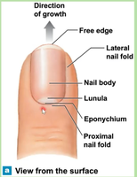
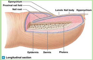
Nails
Nails are protective coverings on the dorsal surfaces of the fingers and toes, composed of keratinized cells.
Nail body: Visible part of the nail.
Nail bed: Skin beneath the nail body.
Nail root: Site of nail production, located deep in the dermis near the bone.
Lunula: Crescent-shaped area at the base of the nail.
Hyponychium: Skin under the free edge of the nail.
Eponychium: Cuticle; protects the area between the skin and the nail from infection.
Summary Table: Layers and Structures of the Skin
Layer/Structure | Main Components | Functions |
|---|---|---|
Epidermis | Keratinoctyes, melanocytes, Merkel cells, Langerhans cells | Protection, water retention, vitamin D3 synthesis, sensation |
Dermis (Papillary) | Loose connective tissue, capillaries, nerves | Nourishes epidermis, sensory input |
Dermis (Reticular) | Dense connective tissue, blood vessels, glands, hair follicles | Strength, elasticity, houses accessory structures |
Hypodermis | Adipose tissue, large blood vessels | Insulation, energy storage, anchors skin |
Accessory Structures | Hair, nails, glands | Protection, sensation, thermoregulation, excretion |
Additional info:
The integumentary system is also involved in social and sexual communication (e.g., pheromones from apocrine glands).
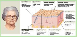
Skin aging leads to reduced repair, decreased immunity, and changes in glandular activity (see image_4 for age-related changes).