 Back
BackThe Integumentary System: Structure, Function, and Clinical Aspects
Study Guide - Smart Notes
 Tailored notes based on your materials, expanded with key definitions, examples, and context.
The Integumentary System
Overview
The integumentary system is the body's largest organ system, comprising the skin and its derivatives. It serves as a protective barrier and plays vital roles in sensation, thermoregulation, and metabolic processes.
Main components: Skin, hair, nails, sweat glands, sebaceous (oil) glands
Functions: Protection, regulation of body temperature, sensation, metabolic functions, excretion
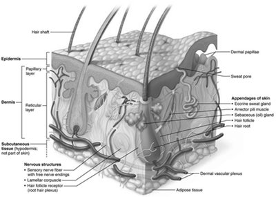
Structure of the Skin
Skin Layers
The skin consists of two main layers and an associated subcutaneous layer:
Epidermis: Superficial, avascular layer made of keratinized stratified squamous epithelium
Dermis: Deeper, vascular layer composed mainly of connective tissue
Hypodermis (subcutaneous layer): Not part of the skin proper; consists mostly of adipose tissue, providing insulation and anchoring skin to underlying structures
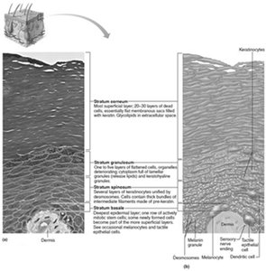
Cells of the Epidermis
Keratinocytes: Produce keratin, a protein that provides protective properties; most abundant cell type in the epidermis
Melanocytes: Produce melanin pigment, which protects against UV radiation
Dendritic (Langerhans) cells: Immune cells that patrol the epidermis
Tactile (Merkel) cells: Sensory receptors for touch
Layers of the Epidermis
The epidermis is organized into distinct layers (strata):
Stratum basale: Deepest layer; single row of mitotically active stem cells and melanocytes
Stratum spinosum: Several layers thick; contains keratinocytes, melanosomes, and dendritic cells
Stratum granulosum: 4–6 layers of flattened cells; keratinization begins, and glycolipids are released for waterproofing
Stratum lucidum: Present only in thick skin (palms, soles); thin, clear layer of dead keratinocytes
Stratum corneum: 20–30 layers of dead, keratinized cells; provides a durable overcoat
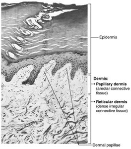
Dermis
The dermis is a strong, flexible connective tissue layer containing nerves, blood vessels, lymphatics, hair follicles, and glands. It is divided into two layers:
Papillary layer: Superficial areolar connective tissue with dermal papillae (fingerlike projections containing capillaries and sensory receptors)
Reticular layer: Deeper, dense irregular connective tissue with collagen and elastic fibers for strength and elasticity
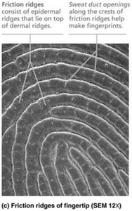
Dermal Modifications and Skin Markings
Friction ridges: Formed by dermal and epidermal ridges; enhance grip and create fingerprints
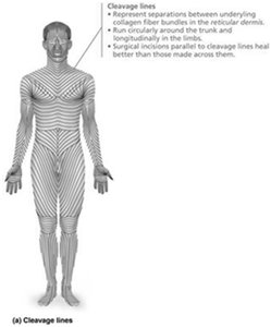
Cleavage lines: Patterns of collagen fibers in the dermis; important for surgical incisions
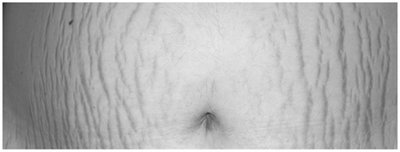
Stretch marks (striae): Result from dermal tearing due to rapid stretching
Skin Color
Pigments Contributing to Skin Color
Melanin: Produced by melanocytes; protects DNA from UV damage; responsible for brown to black skin tones
Carotene: Yellow to orange pigment, most visible in palms and soles; can be converted to vitamin A
Hemoglobin: Oxygenated pigment in red blood cells; gives fair skin a pinkish hue
Alterations in skin color can indicate disease (e.g., cyanosis, pallor, jaundice, erythema, bruises).
Hair
Structure and Function
Hair (pili): Flexible strands of dead, keratinized cells produced by hair follicles
Regions: Shaft (above skin), root (within skin)
Functions: Sensory detection, protection from trauma, heat loss, and sunlight
Hair thinning (alopecia) and baldness (often genetic) can occur with age or hormonal changes.
Nails
Structure and Clinical Significance
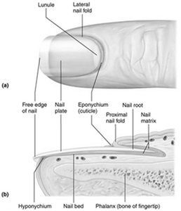
Nails: Scale-like modifications of the epidermis containing hard keratin; protect distal phalanges
Main parts: Free edge, nail plate, root, nail bed, nail matrix (growth area), nail folds, eponychium (cuticle), hyponychium
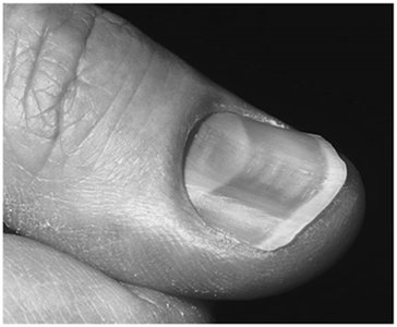
Clinical relevance: Nail color and shape can indicate systemic diseases (e.g., spoon nails in iron deficiency, yellow nails in respiratory disorders)
Glands of the Skin
Types and Functions
Sweat (sudoriferous) glands: Eccrine (merocrine) for thermoregulation; apocrine for scent (axillary/anogenital areas)
Modified apocrine glands: Ceruminous (earwax), mammary (milk)
Sebaceous (oil) glands: Secrete sebum into hair follicles; lubricate skin/hair, antibacterial properties
Gland Type | Function | Secretion | Location |
|---|---|---|---|
Eccrine Sweat | Temperature control, antibacterial | Hypotonic filtrate of plasma | Palms, soles, forehead |
Apocrine Sweat | May act as scent gland | Filtrate with proteins/lipids | Axillary, anogenital |
Sebaceous | Lubricate, antibacterial | Sebum (oil) | Everywhere except palms/soles |
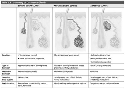
Clinical Correlations
Acne: Inflammation of sebaceous glands, often due to bacterial infection
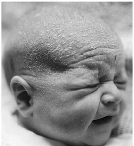
Seborrhea (cradle cap): Overactive sebaceous glands in infants
Functions of the Skin
Protective Barriers
Chemical barrier: Acid mantle, antimicrobial proteins, melanin
Physical barrier: Keratinized cells and glycolipids block water and many substances
Biological barrier: Dendritic cells and macrophages provide immune defense
Other Functions
Body temperature regulation: Sweat and blood flow adjustments
Cutaneous sensations: Touch, pressure, pain, temperature
Metabolic functions: Vitamin D synthesis, hormone activation, collagenase production
Blood reservoir: Holds up to 5% of blood volume
Excretion: Removal of nitrogenous wastes via sweat
Skin Disorders: Cancer and Burns
Skin Cancer
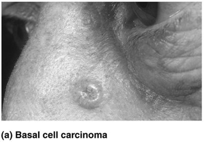
Basal cell carcinoma: Most common, least malignant; arises from stratum basale
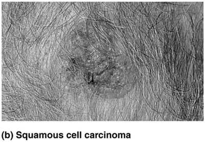
Squamous cell carcinoma: Second most common; arises from keratinocytes of stratum spinosum
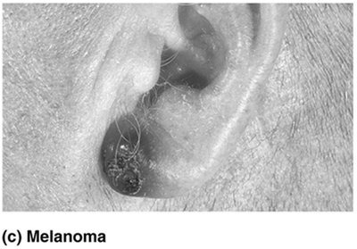
Melanoma: Most dangerous; cancer of melanocytes, highly metastatic
ABCD rule for melanoma detection: Asymmetry, Border irregularity, Color variation, Diameter >6 mm
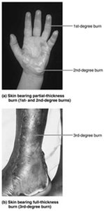
Burns
First-degree: Epidermal damage only; redness, pain
Second-degree: Epidermal and upper dermal damage; blisters
Third-degree: Full-thickness; gray-white, red, or blackened skin, no pain (nerve destruction), requires grafting
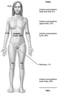
Rule of Nines: Used to estimate burn extent and fluid loss; body divided into sections, each representing 9% (or multiples) of total body surface area.
Developmental and Aging Aspects
Aging skin: Thinner, drier, less elastic; increased risk of injury and cancer
Prevention: UV protection, good nutrition, hydration, hygiene
