Back
BackThe Integumentary System: Structure, Function, and Clinical Relevance
Study Guide - Smart Notes
 Tailored notes based on your materials, expanded with key definitions, examples, and context.
The Integumentary System
Overview and Functions
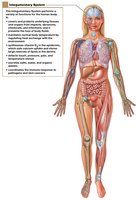
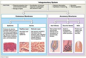
The integumentary system is the largest organ system of the body, comprising the skin and its accessory structures. It serves as the primary barrier between the internal environment and the external world, providing protection, sensory input, and regulatory functions.
Protection: Shields underlying tissues from mechanical damage, pathogens, and chemical exposure.
Excretion: Removes salts, water, and organic wastes through glandular secretions.
Thermoregulation: Maintains body temperature via insulation and evaporative cooling.
Vitamin D Synthesis: Initiates the production of vitamin D3 when exposed to sunlight.
Sensory Reception: Detects touch, pressure, pain, and temperature changes.
Immune Defense: Coordinates immune responses to pathogens and cancers in the skin.
Lipid Storage: Stores lipids in the dermis and subcutaneous layer.
Structure of the Skin
Layers of the Skin
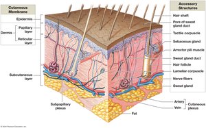
The skin consists of three main layers: the epidermis, dermis, and subcutaneous layer (hypodermis). Each layer has distinct structures and functions.
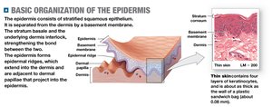
Epidermis: The outermost layer, composed of stratified squamous epithelium. It is avascular and relies on diffusion from the dermis for nutrients.
Dermis: The middle layer, made of connective tissue, containing blood vessels, nerves, and accessory structures.
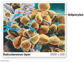
Subcutaneous Layer (Hypodermis): A layer of loose connective and adipose tissue beneath the dermis, providing insulation, energy storage, and anchorage to underlying tissues.
Epidermis
Strata of the Epidermis
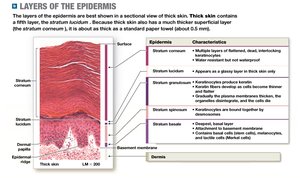
The epidermis is organized into distinct layers (strata), each with specialized cells and functions. There are two types of skin: thin (four layers) and thick (five layers, found on palms and soles).
Stratum | Characteristics |
|---|---|
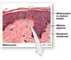
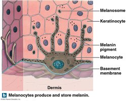
Stratum basale | Deepest layer; contains stem cells (basal cells), melanocytes, and tactile cells; attached to basement membrane. |
Stratum spinosum | 8–10 layers of keratinocytes; contains dendritic (Langerhans) cells for immune defense. |
Stratum granulosum | 3–5 layers; keratinocytes stop dividing, fill with keratin and keratohyalin, and begin to die. |
Stratum lucidum | Present only in thick skin; layer of dead keratinocytes. |
Stratum corneum | 15–30 layers of dead, keratinized cells; provides a tough, water-resistant barrier. |
Specialized Cells of the Epidermis
Keratinocytes: Main cell type, produce keratin for strength and water resistance.
Melanocytes: Produce melanin pigment, protecting against UV radiation.
Tactile (Merkel) Cells: Sensory receptors for touch, found in hairless skin.
Dendritic (Langerhans) Cells: Immune cells that defend against pathogens and cancer.
Epidermal Growth and Water Loss
Keratinization: The process by which cells fill with keratin and move toward the surface, eventually being shed.
Insensible Perspiration: Water loss through diffusion across the stratum corneum (~500 mL/day).
Sensible Perspiration: Water loss via sweat glands, important for thermoregulation.
Epidermal Growth Factor (EGF): Stimulates cell division, keratin production, and repair.
Dermis
Layers of the Dermis
The dermis provides structural support and houses blood vessels, nerves, and accessory structures.
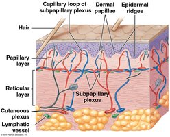
Papillary Layer: Superficial, areolar connective tissue; contains capillaries, lymphatics, and sensory neurons; forms dermal papillae.
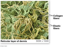
Reticular Layer: Deep, dense irregular connective tissue; rich in collagen and elastic fibers, providing strength and elasticity.
Dermal Features and Blood Supply
Skin Turgor: The skin's flexibility and resilience, dependent on water content.
Tension Lines: Patterns of collagen fibers; incisions parallel to these lines heal better.
Blood Supply: Cutaneous and subpapillary plexuses supply nutrients and aid thermoregulation.
Sensory Receptors: Tactile (Meissner) corpuscles detect light touch; lamellar (Pacinian) corpuscles detect deep pressure and vibration.
Subcutaneous Layer (Hypodermis)
Structure and Function
The subcutaneous layer is not technically part of the skin but is essential for connecting the skin to underlying tissues and providing insulation and energy storage.
Composition: Primarily adipose tissue (adipocytes) and loose connective tissue.
Functions: Insulation, energy storage, shock absorption, and a site for subcutaneous injections.
Skin Color
Pigments and Circulation
Carotene: Orange-yellow pigment from diet, accumulates in epidermal cells and can be converted to vitamin A.
Melanin: Brown-black (eumelanin) or red-yellow (pheomelanin) pigment produced by melanocytes; protects against UV damage.
Hemoglobin: Oxygenated blood gives skin a pinkish hue; low oxygen causes cyanosis (bluish color).
Clinical Conditions Affecting Skin Color
Jaundice: Yellowing due to bilirubin accumulation from liver dysfunction.
Pituitary Tumors/Addison’s Disease: Excess MSH or ACTH causes skin darkening.
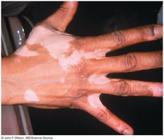
Vitiligo: Autoimmune loss of melanocytes, resulting in white patches.
Vitamin D3 Synthesis
Sunlight and Hormonal Conversion
Exposure to UV radiation in sunlight triggers the synthesis of vitamin D3 (cholecalciferol) in the epidermis. The liver and kidneys convert it to calcitriol, which is essential for calcium and phosphate absorption in the intestine.
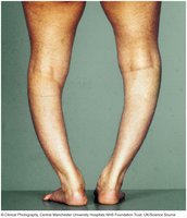
Deficiency: Leads to rickets, characterized by weak, deformed bones.

Hair
Structure and Function
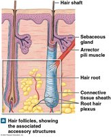
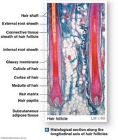
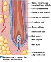
Hair is a nonliving structure produced by hair follicles. It serves protective, sensory, and thermoregulatory roles.
Regions: Hair root (embedded in skin), hair shaft (visible part).
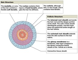
Layers: Medulla (core), cortex (middle), cuticle (outer layer).
Follicle Structure: Internal and external root sheaths, glassy membrane, connective tissue sheath.
Arrector Pili Muscle: Smooth muscle causing "goose bumps."
Hair Growth and Types
Growth Cycle: Active growth, club hair formation, shedding.
Types: Vellus (fine, unpigmented), terminal (coarse, pigmented).
Color: Determined by melanocyte activity and genetics.
Exocrine Glands of the Skin
Types and Secretions
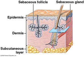
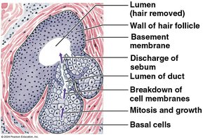
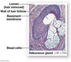
Sebaceous Glands: Secrete sebum (oil) into hair follicles or directly onto skin; lubricates and protects.
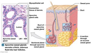
Apocrine Sweat Glands: Secrete sticky, odorous sweat into hair follicles; active after puberty.
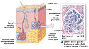
Eccrine (Merocrine) Sweat Glands: Widely distributed; secrete watery sweat directly onto skin for cooling and excretion.
Other Glands: Mammary (milk), ceruminous (earwax).
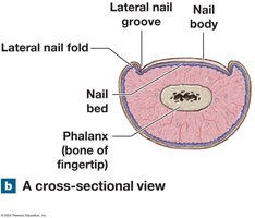
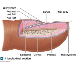
Nails
Structure and Function
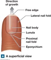
Nails are protective structures composed of keratinized epidermal cells, covering the dorsal surfaces of fingers and toes.
Nail Body: Visible part covering the nail bed.
Nail Root: Site of nail production, hidden under the proximal nail fold.
Lunula: Pale crescent at the base of the nail.
Hyponychium: Thickened stratum corneum beneath the free edge.
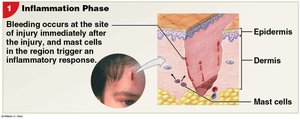
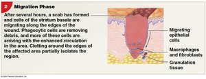
Skin Injury and Repair
Phases of Repair
Inflammatory Phase: Bleeding and inflammation occur; mast cells trigger immune response.
Migration Phase: Scab forms; epithelial cells migrate; macrophages and fibroblasts clean and repair tissue.
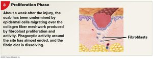
Proliferation Phase: Scab dissolves; fibroblasts produce collagen; epidermal cells cover wound.
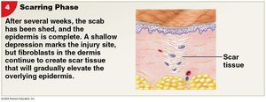
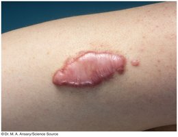
Scarring Phase: Scar tissue forms; may be less flexible and have fewer blood vessels.
Clinical Notes
Burns and Grafts
First-degree: Only epidermis; redness and minor pain.
Second-degree: Epidermis and part of dermis; blistering and pain.
Third-degree: Full thickness; destroys all skin layers; requires grafting.
Rule of Nines: Used to estimate burn surface area for treatment.
Aging and the Integumentary System
Effects of Aging
Epidermal thinning and weakened connections between layers.
Decreased immune function due to fewer dendritic cells.
Reduced vitamin D production and melanocyte activity.
Decline in glandular activity and blood supply, leading to dry skin and impaired thermoregulation.
Hair loss, graying, and slower repair rates.