Back
Ch. 5 - The Integumentary System: Structure, Function, and Clinical Aspects
Study Guide - Smart Notes
 Tailored notes based on your materials, expanded with key definitions, examples, and context.
The Integumentary System
Overview and Structure
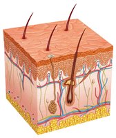
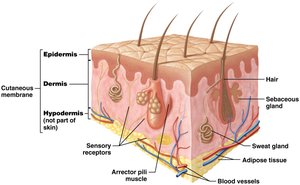
The integumentary system, primarily composed of the skin, is the largest organ system of the human body. It serves as a protective barrier and is involved in various physiological processes. The skin consists of three main layers: the epidermis, dermis, and hypodermis (subcutaneous tissue). Accessory structures such as hair, glands, and nails are also part of this system.
Epidermis: The outermost, avascular layer made of keratinized stratified squamous epithelium.
Dermis: Located beneath the epidermis, composed of loose and dense irregular connective tissue, containing blood vessels, nerves, and sensory receptors.
Hypodermis: Not technically part of the skin, but anchors it to underlying tissues; composed of loose connective and adipose tissue.
Accessory Structures: Hair, glands (sebaceous and sweat), and nails, all derived from keratinized cells.
Functions of the Integument
The integumentary system performs several vital functions essential for homeostasis and survival:
Protection: Acts as a physical barrier against mechanical injury, pathogens, and harmful substances. Melanin protects against UV radiation.
Sensation: Contains sensory receptors that detect changes in the environment (touch, temperature, pain).
Thermoregulation: Regulates body temperature through sweat production and blood vessel dilation/constriction.
Vitamin D Synthesis: Epidermal cells synthesize vitamin D precursor when exposed to UV light.
Excretion: Minor role in excreting waste products through sweat.
Thermoregulation
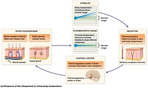
Thermoregulation is controlled by negative feedback loops involving sensory receptors, the hypothalamus, and effectors such as sweat glands and blood vessels. When body temperature rises, sweat glands secrete sweat and blood vessels dilate to dissipate heat. If temperature drops, blood vessels constrict and sweat production decreases.
Microscopic Structure of the Skin
Epidermis: Layers and Cells
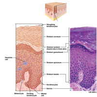
The epidermis is composed of five distinct layers (strata) in thick skin and four in thin skin. The primary cell type is the keratinocyte, but other cells include dendritic (Langerhans) cells, Merkel cells, and melanocytes.
Stratum basale: Single layer of stem cells; site of mitosis and melanin synthesis.
Stratum spinosum: Several layers; limited mitotic activity; contains dendritic cells.
Stratum granulosum: Cells contain keratin and lipid granules; cells begin to die.
Stratum lucidum: Thin, clear layer of dead keratinocytes; present only in thick skin.
Stratum corneum: Multiple layers of dead, keratinized cells; regularly shed and replaced.
Keratinocyte Life Cycle
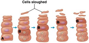
Keratinocytes originate in the stratum basale and migrate upward, undergoing differentiation and eventually dying. The journey from the deepest layer to the surface takes 40–50 days, after which cells are sloughed off.
Thick vs. Thin Skin
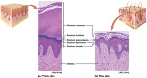
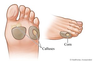
Thick skin (palms, soles) contains all five strata and a thick stratum corneum, but lacks hair follicles. Thin skin (most of the body) lacks the stratum lucidum, has a thinner stratum corneum, and contains hair follicles and sebaceous glands. Calluses form in response to repeated friction or pressure.
The Dermis
Structure and Layers
The dermis is a highly vascular connective tissue layer beneath the epidermis, providing structural support and nourishment. It consists of two layers:
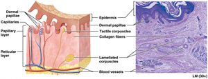
Papillary layer: Superficial, composed of loose connective tissue; contains dermal papillae, capillaries, and tactile (Meissner) corpuscles for touch sensation.
Reticular layer: Deeper, composed of dense irregular connective tissue; contains collagen and elastic fibers, lamellated (Pacinian) corpuscles for pressure/vibration, blood vessels, glands, and hair follicles.
Skin Markings
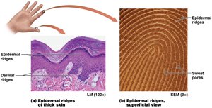
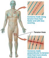
Skin markings such as epidermal ridges (fingerprints) are formed by the interaction of the dermis and epidermis, enhancing grip. Tension (cleavage) lines correspond to the orientation of collagen bundles and are important in surgical incisions. Flexure lines occur where the skin is tightly bound to underlying structures.
Skin Color
Pigments and Determinants
Skin color is determined by three main pigments:
Melanin: Orange-red to black pigment produced by melanocytes; protects against UV radiation.
Carotene: Yellow-orange pigment from diet; accumulates in the stratum corneum.
Hemoglobin: Red pigment in blood; imparts a reddish hue depending on blood flow and oxygenation.
Melanin Production and Function
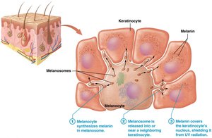
Melanin is synthesized from tyrosine by the enzyme tyrosinase in melanosomes. Melanin is transferred to keratinocytes, where it forms a protective cap over the nucleus to shield DNA from UV damage. Melanin production increases with UV exposure, resulting in tanning. It also reduces vitamin D synthesis, influencing evolutionary adaptations in skin color.
Clinical Relevance of Skin Color
Skin color can indicate underlying health conditions:
Freckles: Increased melanin production in localized areas.
Moles (nevi): Increased number of melanocytes.
Albinism: Lack of melanin production.
Hypercarotenemia: Excess carotene in the blood.
Erythema: Redness due to increased blood flow.
Pallor: Paleness due to decreased blood flow.
Cyanosis: Bluish tint due to low oxygenation of hemoglobin.

Accessory Structures
Hair (Pili)
Hair covers most of the body except areas of thick skin, lips, and parts of the external genitalia. It provides limited thermoregulation, protects against UV radiation, and prevents entry of foreign particles. Hair is composed of dead, keratinized cells and is associated with sensory neurons.
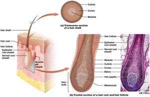

Hair Structure and Growth
Each hair consists of a shaft (above the skin) and a root (embedded in the skin). The hair bulb contains the matrix, where growth occurs. The arrector pili muscle attaches to the follicle, causing piloerection (goosebumps). Hair growth occurs in cycles of active growth and resting phases.
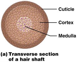
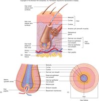
Hair Types and Pigmentation
Hair types include lanugo (fetal), terminal (thick, pigmented), and vellus (thin, nonpigmented). Hair color is determined by the type and amount of melanin produced by melanocytes. With age, melanin production decreases, leading to gray or white hair.
Glands of the Integument
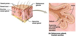
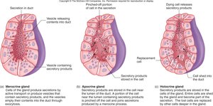
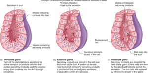
Skin glands are derived from the epidermis but located in the dermis. The two main types are sebaceous (oil) glands and sudoriferous (sweat) glands.
Sebaceous glands: Secrete sebum, an oily substance that lubricates and waterproofs skin and hair; secretion increases with sex hormones.
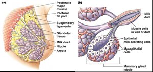
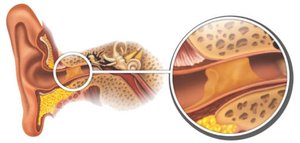
Sudoriferous glands: Four types—eccrine (most abundant, watery sweat), apocrine (protein-rich, odoriferous sweat), ceruminous (ear wax), and mammary (milk production).

Pathophysiology of the Integument
Burns
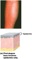
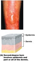
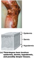
Burns are classified by depth and extent of tissue damage:
First-degree: Damage to epidermis only; redness and mild pain.
Second-degree: Damage to epidermis and dermis; blistering and scarring.
Third-degree: Damage extends to hypodermis or deeper; risk of infection, scarring, and loss of accessory structures.
Rule of Nines
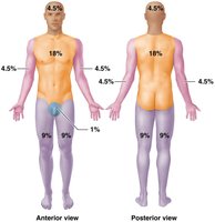
The Rule of Nines divides the body into regions representing 9% (or multiples) of total body surface area, aiding in the assessment and treatment of burns.
Skin Cancer
Skin cancer arises from mutations in skin cells, often due to UV exposure. The three main types are:
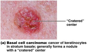
Basal cell carcinoma: Most common; arises from stratum basale; rarely metastasizes.
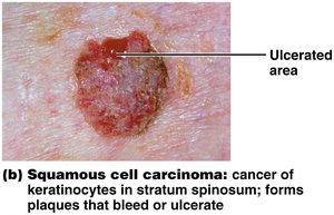
Squamous cell carcinoma: Arises from stratum spinosum; may metastasize.
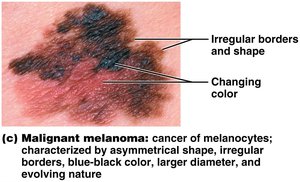
Malignant melanoma: Cancer of melanocytes; highly metastatic and dangerous.
Effects of Aging on the Integument
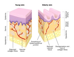
Aging causes thinning of the epidermis, decreased collagen, reduced elasticity, increased dryness, impaired thermoregulation, and altered melanin function, leading to increased susceptibility to injury, infection, and wrinkling.