 Back
The Integumentary System: Structure, Function, and Clinical Aspects
Study Guide - Smart Notes
 Tailored notes based on your materials, expanded with key definitions, examples, and context.
Chapter 5: The Integumentary System
Overview of the Integumentary System
The integumentary system is composed of the skin and its derivatives (hair, nails, glands). It serves as the body's first line of defense and plays critical roles in protection, sensation, thermoregulation, and metabolic functions.
Skin Structure
Layers of the Skin
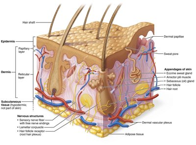
Epidermis: The outermost, avascular layer composed of keratinized stratified squamous epithelium. It provides a waterproof barrier and creates our skin tone.
Dermis: The middle layer, made of strong, flexible connective tissue. It contains blood vessels, nerves, and appendages of the skin.
Hypodermis (Subcutaneous Tissue): Not part of the skin proper, but lies below the dermis. It consists mainly of adipose tissue and anchors skin to underlying structures.
Cells of the Epidermis
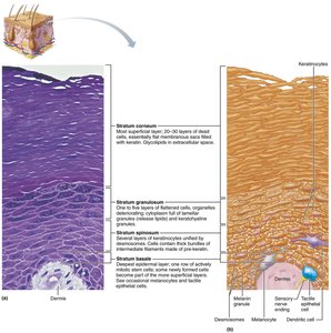
Keratinocytes: The majority of epidermal cells; produce keratin, a tough, fibrous protein that provides protection.
Melanocytes: Synthesize melanin, the pigment that protects the nucleus from UV radiation. Found in the stratum basale.
Dendritic (Langerhans) Cells: Immune cells that ingest foreign substances and activate the immune system.
Tactile (Merkel) Cells: Associated with sensory nerve endings; function as touch receptors.
Layers of the Epidermis
The epidermis is organized into distinct layers (from deep to superficial):
Stratum Basale: Deepest layer; contains stem cells that divide to form new keratinocytes.
Stratum Spinosum: Several layers of keratinocytes unified by desmosomes; contains dendritic cells.
Stratum Granulosum: Cells flatten, organelles deteriorate, and keratinization begins; contains lamellar granules for waterproofing.
Stratum Lucidum: Present only in thick skin (palms, soles); a thin, translucent band of dead keratinocytes.
Stratum Corneum: Outermost layer; 20–30 layers of dead, keratinized cells that provide a durable overcoat.
The Dermis
Structure and Function
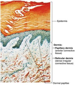
The dermis is a connective tissue layer supporting the epidermis and housing blood vessels, nerves, and skin appendages.
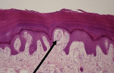
Papillary Layer: Superficial areolar connective tissue with dermal papillae that project into the epidermis, containing capillaries, nerve endings, and Meissner's corpuscles (touch receptors).
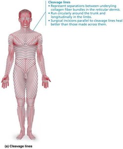
Reticular Layer: Deeper, dense irregular connective tissue; provides strength and elasticity, contains collagen fibers, and forms cleavage (tension) lines important for surgical incisions.
Dermal Specializations
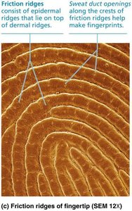
Friction Ridges: Found in thick skin; enhance grip and form fingerprints due to sweat duct openings along the ridges.
Cleavage (Tension) Lines: Externally invisible lines representing the orientation of collagen fibers; incisions parallel to these lines heal better.
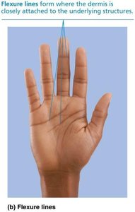
Flexure Lines: Dermal folds at or near joints where the skin is tightly bound to underlying structures, visible on palms and fingers.
Clinical Correlates
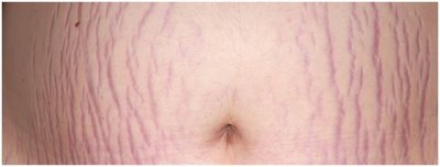
Striae (Stretch Marks): Dermal tears from extreme stretching, appearing as silvery white scars.
Blisters: Fluid-filled pockets separating the epidermal and dermal layers due to acute trauma.
Skin Color
Pigments Contributing to Skin Color
Melanin: Only pigment produced in the skin; ranges from reddish-yellow to brownish-black. Increased by sun exposure for UV protection.
Carotene: Yellow to orange pigment, most visible in the palms and soles; accumulates in the stratum corneum and hypodermis.
Hemoglobin: Oxygenated hemoglobin in dermal capillaries gives fair skin a pinkish hue.
Alterations in skin color can indicate disease (e.g., cyanosis, pallor, erythema, jaundice, bruises, and hyperpigmentation in endocrine disorders).
Hair
Structure and Function of Hair
Hair consists of dead, keratinized cells and is found almost everywhere except palms, soles, lips, nipples, and parts of external genitalia. Its main functions include sensory input, protection from trauma, and filtration of particles.
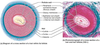
Shaft: Visible part above the scalp; keratinization is complete.
Root: Part within the scalp; keratinization is ongoing.
Medulla: Central core with large cells and air spaces.
Cortex: Several layers of flattened cells surrounding the medulla.
Cuticle: Outermost layer of overlapping single cells.
Hair color is determined by melanins produced in hair follicles; gray/white hair results from decreased melanin and air bubbles in the shaft.
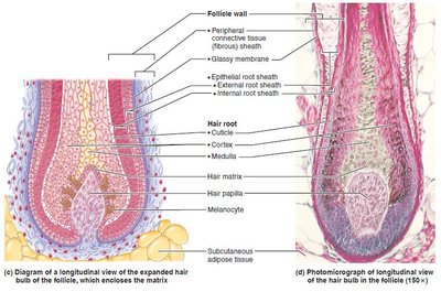
Hair Follicle Structure
Hair Bulb: Expanded deep end of the follicle; contains the hair matrix (actively dividing cells).
Hair Papilla: Dermal tissue with capillaries supplying nutrients.
Arrector Pili: Smooth muscle causing "goose bumps."
Root Hair Plexus: Sensory nerve endings around the bulb, making hair a touch receptor.
Types and Growth of Hair
Vellus Hair: Pale, fine body hair of children and adult females.
Terminal Hair: Coarse, long hair of scalp, eyebrows, and, after puberty, axillary and pubic regions.
Hair growth is influenced by nutrition and hormones; follicles cycle between active and regressive phases.
Clinical Correlates: Hair Disorders
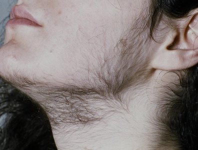
Hirsutism: Excessive hairiness due to increased androgens, often from ovarian or adrenal tumors.
Alopecia: Hair thinning with age.
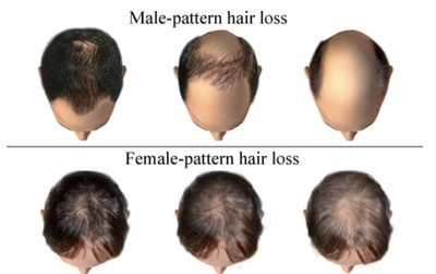
Male Pattern Baldness: Genetically determined, sex-influenced, and related to follicular response to DHT (dihydrotestosterone).
Nails
Structure and Function
Nails are scale-like modifications of the epidermis containing hard keratin. They protect the distal, dorsal surfaces of fingers and toes and aid in picking up small objects.
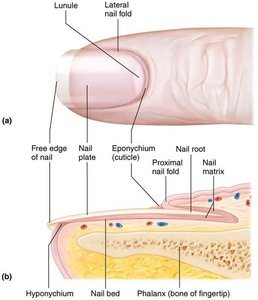
Free Edge: The part that extends beyond the finger or toe.
Nail Plate: The visible attached portion.
Nail Root: Embedded in the skin.
Nail Matrix: Thickened proximal portion responsible for nail growth.
Eponychium (Cuticle): Proximal nail fold projecting onto the nail body.
Hyponychium: Area under the free edge that accumulates dirt.
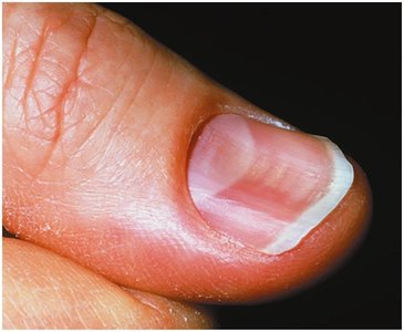
Clinical Correlates: Nail Disorders
Yellow Nails: May indicate respiratory or thyroid disorders.
Thickened Yellow Nails: Often due to fungal infection.
Koilonychia (Spoon Nail): Outward concavity, may signal iron deficiency.
Beau’s Lines: Horizontal lines indicating severe illness.
Cutaneous Glands
Sweat (Sudoriferous) Glands
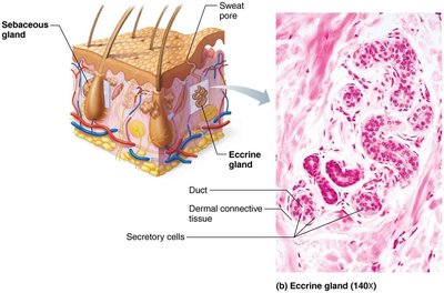
Eccrine (Merocrine) Glands: Most numerous; abundant on palms, soles, and forehead. Secrete sweat for thermoregulation.
Apocrine Glands: Confined to axillary and anogenital areas; secrete viscous, milky sweat. Begin functioning at puberty and may act as scent glands.
Modified Apocrine Glands: Ceruminous glands (earwax) and mammary glands (milk).
Sebaceous (Oil) Glands
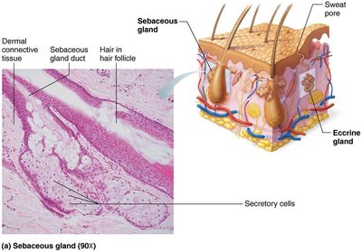
Widely distributed except in thick skin of palms and soles.
Secrete sebum (oily holocrine secretion) into hair follicles; lubricates and softens hair and skin, has bactericidal properties.
Activity increases at puberty due to androgens.
Summary Table: Cutaneous Glands
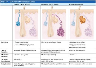
Gland Type | Function | Secretion | Method of Secretion | Location |
|---|---|---|---|---|
Eccrine Sweat Glands | Temperature control, antibacterial properties | Hypotonic filtrate of blood plasma | Merocrine (exocytosis) | Everywhere, especially palms, soles, forehead |
Apocrine Sweat Glands | May act as sexual scent glands | Filtrate of blood plasma with added proteins and fatty substances | Merocrine (exocytosis) | Axillary and anogenital regions |
Sebaceous Glands | Lubricate skin and hair, antibacterial properties | Sebum (oil) | Holocrine | Everywhere except palms and soles |
Clinical Correlates: Gland Disorders
Acne: Inflammatory disease of sebaceous glands, often due to bacterial infection.
Whiteheads/Blackheads: Blocked sebaceous glands; oxidation turns whiteheads into blackheads.
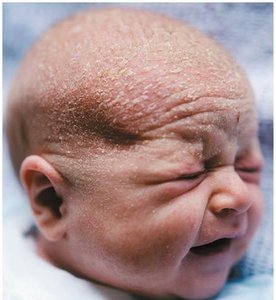
Seborrhea (Cradle Cap): Overactive sebaceous glands in infants, causing flaky scalp lesions.
Functions of the Skin
Protection
Chemical Barrier: Skin secretions (sweat, sebum) and melanin protect against microbes and UV radiation.
Physical Barrier: Keratinized cells block water and most substances.
Biological Barrier: Dendritic cells and macrophages provide immune defense.
Body Temperature Regulation
Insensible perspiration (500 ml/day) occurs under normal conditions.
Sensible perspiration (up to 12 L/day) occurs when body temperature rises, aiding cooling.
Constriction of dermal blood vessels conserves heat in cold environments.
Cutaneous Sensations
Receptors for touch, temperature, and pain are present in the skin.
Meissner’s corpuscles detect light touch; free nerve endings sense pain.
Metabolic Functions
Synthesizes vitamin D for calcium absorption.
Keratinocytes activate hormones and disarm carcinogens.
Produces collagenase for collagen turnover.
Blood Reservoir and Excretion
Holds up to 5% of total blood volume; can shunt blood to other organs.
Excretes nitrogenous wastes and participates in salt/water balance via sweat.
Skin Cancer
Types and Characteristics
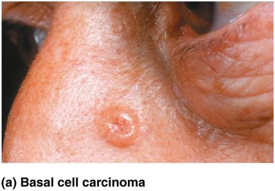
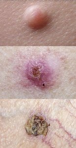
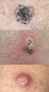
Basal Cell Carcinoma: Most common, least malignant; arises from stratum basale. Cured by surgical excision in most cases.
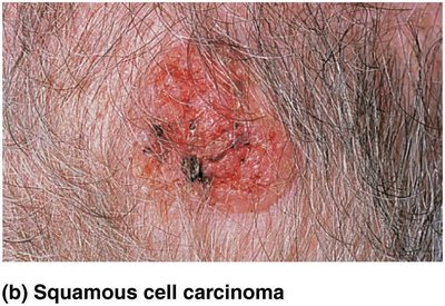
Squamous Cell Carcinoma: Second most common; arises from keratinocytes of stratum spinosum. Can metastasize but has a good prognosis if treated early.
Melanoma: Cancer of melanocytes; most dangerous due to high metastatic potential and resistance to chemotherapy. Early detection is critical (ABCD rule: Asymmetry, Border, Color, Diameter, Evolution).
Burns
Classification and Clinical Importance
First-Degree Burns: Affect only the epidermis; cause redness, swelling, and pain.
Second-Degree Burns: Affect the epidermis and upper dermis; blisters appear.
Third-Degree Burns: Involve the entire thickness of the skin; skin appears gray-white, cherry red, or blackened. Not painful due to destroyed nerve endings; requires skin grafting.
Fourth-Degree Burns: Extend into deeper tissues such as muscle and bone.
Burns are considered critical if >25% of body has second-degree burns, >10% has third-degree burns, or if face, hands, or feet are involved. Treatment includes fluid replacement, infection control, and skin grafts.
*Additional info: The "Rule of Nines" is used to estimate the percentage of body surface area affected by burns for fluid resuscitation purposes.*
