 Back
The Integumentary System: Structure, Function, and Clinical Relevance
Study Guide - Smart Notes
 Tailored notes based on your materials, expanded with key definitions, examples, and context.
The Integumentary System
Overview
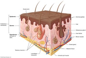
The integumentary system is composed of the skin and its accessory structures, including hair, nails, and glands. It is the largest organ system in the body, vital for protection, sensation, thermoregulation, excretion, and vitamin D synthesis.
Skin (Cutaneous Membrane): Largest organ, 10-15% of body weight.
Two main components:
Superficial Epidermis: Keratinized stratified squamous epithelium.
Deep Dermis: Loose and dense irregular connective tissue.
Accessory structures: Hair, nails, sweat glands, sebaceous glands.
Hypodermis: Not considered part of the skin in this course; consists of loose connective and adipose tissue.
Functions of the Integumentary System
The skin is essential for maintaining homeostasis and performs several key functions:
Protection: Shields underlying tissues from mechanical trauma, pathogens, and environmental threats. The acid mantle (slightly acidic pH) deters pathogen growth.
Sensation: Sensory receptors detect harmful stimuli, crucial for homeostasis.
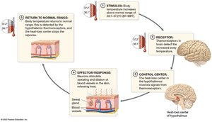
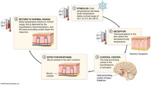
Thermoregulation: Maintains stable internal temperature via negative feedback loops.
When body temperature rises: Thermoreceptors signal the hypothalamus, which triggers sweating and vasodilation to release heat.
When body temperature falls: Thermoreceptors signal the hypothalamus, which triggers vasoconstriction to conserve heat.
Excretion: Eliminates small amounts of metabolic waste (lactic acid, urea) in sweat.
Vitamin D Synthesis: UV exposure converts a precursor to cholecalciferol, which is modified by the liver and kidneys to form calcitriol, essential for calcium absorption.
Structure of the Skin
The Epidermis
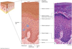
The epidermis is the superficial layer of the skin, composed of several cell types and up to five distinct layers (strata).
Keratinocytes: 95% of epidermal cells; produce keratin for strength and resistance to stress.
Layers (from deep to superficial):
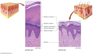
Stratum Basale: Single layer of stem cells; mitotically active; contains melanocytes and Merkel cells.
Stratum Spinosum: Thickest layer; metabolically active; contains dendritic cells.
Stratum Granulosum: 3-5 rows; cells contain granules; release lipids for water barrier; produce filaggrin and acid mantle.
Stratum Lucidum: 2-3 layers; clear, dead keratinocytes; only in thick skin.
Stratum Corneum: 20-30 layers of dead, flattened keratinocytes; cells are shed daily.
Other Cells of the Epidermis
Dendritic (Langerhans) Cells: Immune phagocytes in stratum spinosum.
Tactile (Merkel) Cells: Sensory receptors in stratum basale; detect light touch.
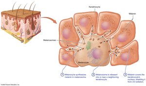
Melanocytes: Produce melanin in stratum basale.
Thick and Thin Skin
Thick Skin: Palms, soles, fingers, toes; all five strata; thick stratum corneum; no hair follicles.
Thin Skin: Covers most of the body; lacks stratum lucidum; thinner stratum corneum; contains hair follicles, sweat, and sebaceous glands.
Callus: Extra layers of stratum corneum from repeated pressure.
The Dermis
The dermis is the deep layer of the skin, providing structural support and housing blood vessels, nerves, and glands.
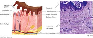
Papillary Layer: Superficial; loose connective tissue; dermal papillae provide blood supply and house tactile corpuscles.
Reticular Layer: Deep; dense irregular connective tissue; contains blood vessels, sweat glands, hair follicles, sebaceous glands, and lamellated corpuscles.
Skin Markings
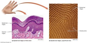
Dermal Ridges: Prominent in thick skin; form epidermal ridges (fingerprints).
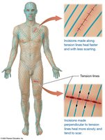
Tension (Cleavage) Lines: Gaps between collagen bundles; important for surgical incisions.
Flexure Lines: Deep creases in areas like palms.
Pigmentation and Skin Color
Melanin
Produced by melanocytes in stratum basale; stored in melanosomes.
Types: Eumelanin (black/brown), Pheomelanin (yellow/red).
Function: Shields DNA from UV radiation; must be replaced regularly.
UV exposure: Increases melanin synthesis; controls vitamin D production.
Common Pigmentation Variations
Freckle: Localized increased melanin production.
Mole (Nevus): Local proliferation of melanocytes.
Albinism: Lack of tyrosinase; no skin pigmentation; increased risk of UV damage.
Other Pigments Affecting Skin Color
Carotene: Yellow-orange pigment from diet; accumulates in stratum corneum.
Hemoglobin: Iron-containing protein in red blood cells; gives skin a pinkish hue in fair-skinned individuals.
Skin Color as a Diagnostic Tool
Erythema: Increased blood flow; red appearance.
Pallor: Decreased blood flow; pale appearance.
Cyanosis: Low oxygen; bluish appearance.
Jaundice: Bilirubin buildup; yellow-orange appearance.
Accessory Structures: Hair, Nails, and Glands
Hair
Structure:
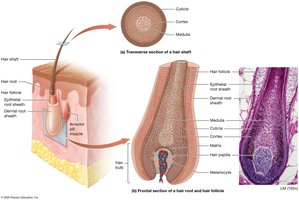
Hair Shaft: Dead keratinized cells; three regions (medulla, cortex, cuticle).
Hair Root: Embedded in dermis; living cells; matrix cells divide for growth.
Hair Follicle: Infolding of epidermis; surrounded by dermal root sheath.
Arrector Pili Muscles: Cause piloerection (goosebumps).
Hair Bulb: Base of root; contains hair papilla (capillaries).
Growth: Alternates between growth and resting stages; alopecia is baldness.
Pigment and Texture: Determined by melanin; changes with age.
Types: Lanugo (fetus), Terminal (scalp, eyes), Vellus (body).
Nails
Structure: Stratified squamous epithelium with hard keratin.
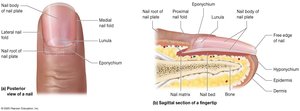
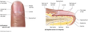
Nail Plate: Sits on nail bed; divided into nail body and nail root.
Nail Matrix: Actively dividing cells under nail root.
Protective Folds: Proximal nail fold, eponychium (cuticle), medial/lateral folds, hyponychium.
Growth: Continuous; lunula is opaque region with accumulated keratin.
Pigment: No melanocytes; color reflects oxygenation.
Glands
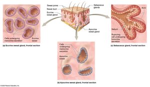
Sweat (Sudoriferous) Glands:
Eccrine: Most prevalent; thermoregulation; watery sweat.
Apocrine: Axillae, anal area, areolae; thick, protein-rich sweat; odor from bacterial metabolism.
Ceruminous: Modified apocrine; produce cerumen (ear wax).
Mammary: Specialized; produce milk.
Sebaceous Glands: Branched; produce sebum via holocrine secretion; increases at puberty; associated with acne.
Acne
Acne Vulgaris: Accumulation of sebum and dead cells in sebaceous glands; can become infected and inflamed.
Male sex hormones: Contribute to acne development.
Skin Cancer
Overview
Skin cancer is a common disease linked to UV exposure and other carcinogens. It results from mutations causing uncontrolled cell growth and tumor formation.
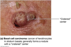
Basal Cell Carcinoma: Most common; arises from stratum basale keratinocytes; rarely metastasizes.
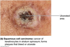
Squamous Cell Carcinoma: Second most common; arises from stratum spinosum keratinocytes; more likely to metastasize.
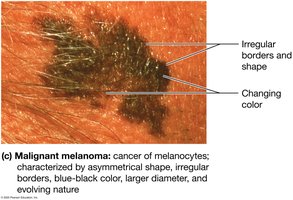
Malignant Melanoma: Least common but most dangerous; arises from melanocytes; high tendency to metastasize.
ABCDE Rule for Melanoma
A: Asymmetrical shape
B: Border irregularity
C: Color (blue-black or varied)
D: Diameter (>6 mm)
E: Evolving shape and size
Summary Table: Layers and Functions of the Skin
Layer | Main Cell Types | Key Functions |
|---|---|---|
Epidermis | Keratinocytes, Melanocytes, Merkel, Langerhans | Protection, Sensation, Vitamin D synthesis |
Dermis | Fibroblasts, Adipocytes, Blood vessels | Support, Thermoregulation, Sensation |
Hypodermis | Adipocytes | Insulation, Energy storage |
Summary Table: Gland Types
Gland Type | Location | Secretion | Function |
|---|---|---|---|
Eccrine | Most skin | Watery sweat | Thermoregulation |
Apocrine | Axillae, anal, areolae | Protein-rich sweat | Odor, sexual signaling |
Ceruminous | Ear canal | Cerumen | Protects eardrum |
Mammary | Breasts | Milk | Nourishes infant |
Sebaceous | Face, scalp | Sebum | Lubricates skin/hair |
