 Back
BackThe Lymphatic and Immune Systems: Structure, Function, and Clinical Relevance
Study Guide - Smart Notes
 Tailored notes based on your materials, expanded with key definitions, examples, and context.
The Lymphatic and Immune Systems
Overview and Learning Outcomes
The lymphatic and immune systems are essential for maintaining fluid balance, defending against pathogens, and supporting overall health. Understanding their structure and function is crucial for students in allied health professions, as these systems are frequently involved in clinical scenarios and patient care.
Build Foundational Anatomical Vocabulary & Concepts: Mastery of anatomical and clinical terms related to the lymphatic and immune systems.
Connect Anatomy and Physiology to Health Careers: Application of knowledge to clinical and practical scenarios.
Develop Clinical Reasoning: Analyze and predict clinical findings related to lymphatic and immune function.
Support Exam Readiness: Objectives and assessments align with board-style and licensure exams.
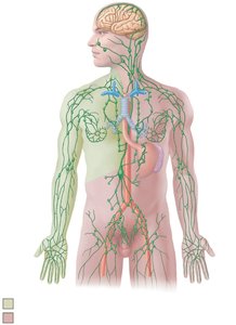
General Structure and Distribution of the Lymphatic System
Major Components and Pathways
The lymphatic system consists of a network of vessels, nodes, and organs that transport lymph and facilitate immune responses. It parallels the venous system and drains excess interstitial fluid back to the bloodstream.
Lymphatic Vessels: Begin as blind-ended capillaries in tissues, merging into larger collecting vessels and ducts.
Lymph Nodes: Small, bean-shaped structures that filter lymph and house immune cells.
Major Ducts: The right lymphatic duct and thoracic duct return lymph to the venous circulation.
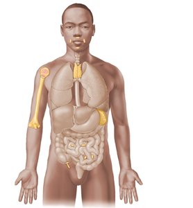
Regional Nodes: Cervical, axillary, and inguinal nodes are key sites for lymph filtration.
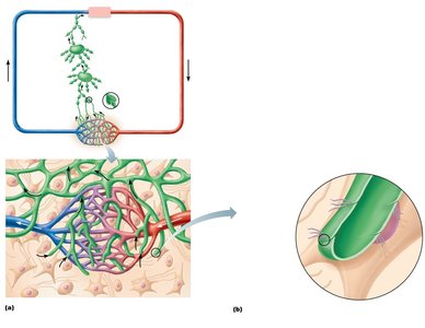
Lymphatic Capillaries and Fluid Flow
Structure and Function of Lymphatic Capillaries
Lymphatic capillaries are highly permeable, blind-ended tubes that collect excess tissue fluid (lymph) and return it to the bloodstream. Their unique structure allows for efficient uptake of fluid, proteins, and particulate matter.
Flaplike Minivalves: Overlapping endothelial cells form one-way valves that open when interstitial pressure increases.
Anchoring Filaments: Attach capillaries to surrounding connective tissue, preventing collapse.
Relationship to Blood Capillaries: Lymphatic capillaries are interwoven with blood capillary beds, facilitating fluid exchange.
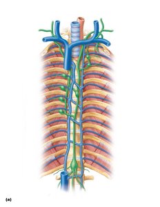
Lymphatic Trunks and Ducts
Major Lymphatic Trunks and Ducts
Lymph from collecting vessels drains into larger lymphatic trunks, which then empty into the right lymphatic duct or thoracic duct. These ducts return lymph to the venous system at the junction of the internal jugular and subclavian veins.
Right Lymphatic Duct: Drains lymph from the right upper limb, right side of the head and thorax.
Thoracic Duct: Drains lymph from the rest of the body; begins at the cisterna chyli.
Major Trunks: Jugular, subclavian, bronchomediastinal, intestinal, and lumbar trunks.
Lymphoid Tissue and Mucosa-Associated Lymphoid Tissue (MALT)
Structure and Function of MALT
Mucosa-associated lymphoid tissue (MALT) consists of lymphoid follicles located in mucous membranes throughout the body, especially in the digestive, respiratory, and genitourinary tracts. MALT provides immune surveillance and response at common entry points for pathogens.
Lymphoid Follicles: Aggregates of lymphocytes, often with germinal centers for B cell proliferation.
Locations: Tonsils, Peyer's patches in the small intestine, and the appendix.
Lymphoid Organs
Primary and Secondary Lymphoid Organs
Lymphoid organs are classified as primary or secondary based on their roles in lymphocyte development and immune response.
Primary Lymphoid Organs: Sites of lymphocyte formation and maturation (red bone marrow and thymus).
Secondary Lymphoid Organs: Sites where mature lymphocytes encounter antigens (lymph nodes, spleen, tonsils, aggregated lymphoid nodules, appendix).
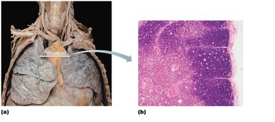
The Thymus
Structure and Function of the Thymus
The thymus is a primary lymphoid organ located in the superior mediastinum. It is essential for T lymphocyte (T cell) maturation and is most active during childhood.
Cortex: Densely packed with immature T cells.
Medulla: Contains mature T cells and thymic corpuscles (Hassall's corpuscles).
Involution: The thymus decreases in size and activity after puberty.
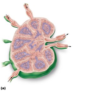
Lymph Nodes
Structure and Function of Lymph Nodes
Lymph nodes are small, encapsulated organs that filter lymph and provide sites for immune cell activation. They are distributed along lymphatic vessels and are especially concentrated in the cervical, axillary, and inguinal regions.
Afferent Vessels: Bring lymph into the node.
Cortex: Contains lymphoid follicles with germinal centers.
Medulla: Contains medullary cords and sinuses.
Efferent Vessels: Carry filtered lymph away from the node.
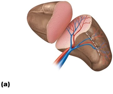
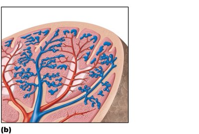
The Spleen
Structure and Function of the Spleen
The spleen is the largest lymphoid organ, located in the upper left abdomen. It filters blood, removes old erythrocytes, and mounts immune responses to blood-borne antigens.
Red Pulp: Contains sinusoids and splenic cords; site of erythrocyte destruction.
White Pulp: Contains lymphocytes arranged around central arteries; site of immune activation.
Hilum: Entry and exit point for splenic vessels.
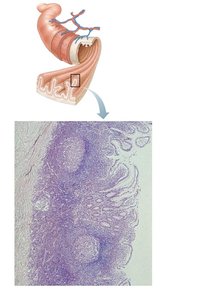
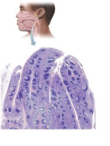
Tonsils and Aggregated Lymphoid Nodules
Structure and Function of Tonsils
Tonsils are collections of lymphoid tissue located in the pharynx. They protect against inhaled or ingested pathogens and contain crypts that trap antigens for immune processing.
Types: Pharyngeal, palatine, and lingual tonsils.
Histology: Lymphoid follicles with germinal centers and crypts.
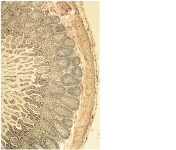
Aggregated Lymphoid Nodules (Peyer's Patches)
Peyer's patches are large clusters of lymphoid follicles found in the wall of the small intestine. They monitor intestinal bacteria and prevent the growth of pathogenic microbes.
Location: Primarily in the ileum of the small intestine.
Function: Initiate immune responses to antigens in the gut.
Clinical Connections and Case Scenarios
Edema and Lymphatic Obstruction
Edema is swelling caused by excess fluid in tissues. It can result from lymphatic vessel blockage or removal, which impairs fluid drainage and leads to accumulation in the interstitial space.
Causes: Infection, surgery, cancer, or congenital defects.
Clinical Relevance: Recognizing and managing edema is important in patient care, especially after lymph node removal or in cases of lymphedema.
Clinical Takeaway
Understanding the lymphatic and immune systems is essential for diagnosing and managing conditions such as infections, autoimmune diseases, allergies, and cancer metastasis. Mastery of these concepts supports success on board exams and in clinical practice.
