 Back
BackThe Lymphatic System and Immunity: Structure and Function
Study Guide - Smart Notes
 Tailored notes based on your materials, expanded with key definitions, examples, and context.
The Lymphatic System and Immunity
Introduction to the Lymphatic System
The lymphatic system is a network of vessels, tissues, and organs that play a crucial role in defending the body against pathogens and maintaining fluid balance. It includes lymphatic vessels, lymph nodes, tonsils, the thymus, spleen, and bone marrow.
Primary Functions: Defense against environmental hazards (e.g., pathogens) and internal threats (e.g., cancer cells).
Main Components: Lymphatic vessels, lymph (fluid), lymphocytes, and lymphoid organs/tissues.
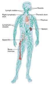
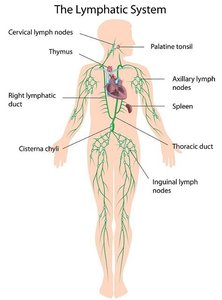
Lymphatic System Anatomy
The lymphatic system consists of a network of lymphatic vessels that transport lymph, a fluid similar to interstitial fluid, throughout the body. Lymphoid organs and tissues are distributed along these vessels and are sites of immune cell production and activation.
Lymphatic Vessels: Begin as lymphatic capillaries in peripheral tissues and merge into larger vessels that return lymph to the venous system.
Lymph Nodes: Small, bean-shaped organs that filter lymph and house immune cells.
Tonsils, Thymus, Spleen, Appendix, and MALT: Specialized lymphoid tissues and organs with immune functions.
Lymphocytes: The Primary Cells of Immunity
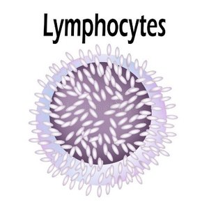
Lymphocytes are the main cellular components of the lymphatic system, responsible for recognizing and responding to specific antigens. They are produced in lymphoid tissues and red bone marrow.
Types of Lymphocytes:
T cells: Responsible for cell-mediated immunity; attack infected or abnormal cells.
B cells: Responsible for antibody-mediated (humoral) immunity; produce antibodies.
NK (Natural Killer) cells: Provide immunological surveillance by attacking foreign, infected, or cancerous cells.
Antigens: Substances that provoke an immune response, usually proteins or other organic molecules.
Lymphatic Capillaries and Vessels
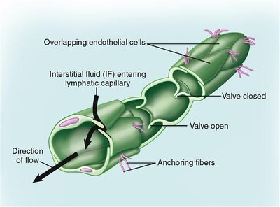
Lymphatic capillaries are the smallest lymphatic vessels, present in almost every tissue. They collect interstitial fluid and transport it as lymph. These capillaries have unique structural features that facilitate their function.
Key Features:
Originate as blind-ended tubes in the periphery.
Have larger diameters and thinner walls than blood capillaries.
Endothelial cells overlap to form one-way valves, allowing fluid and large solutes to enter but not exit.
Major Lymphatic Vessels and Drainage
Larger lymphatic vessels are organized into trunks and ducts that drain lymph from different regions of the body and return it to the venous circulation.
Lymphatic Trunks: Jugular, subclavian, bronchomediastinal, lumbar, and intestinal trunks.
Cisterna Chyli: An expanded chamber receiving lymph from the lumbar and intestinal trunks.
Lymphatic Ducts:
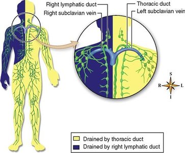
Right Lymphatic Duct: Drains right arm, right upper torso, right head and neck.
Thoracic Duct: Drains the rest of the body.
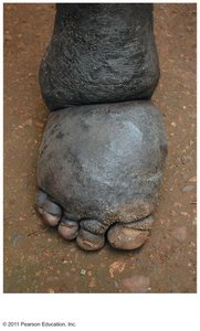
Lymphedema
Lymphedema is a condition resulting from the blockage of lymphatic drainage, leading to the accumulation of interstitial fluid and swelling, most commonly in the limbs. Chronic lymphedema can lead to infection due to stagnant fluid.
Lymphopoiesis: Formation of Lymphocytes
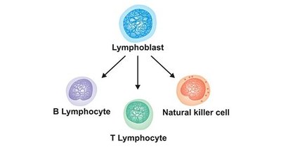
Lymphopoiesis is the process of lymphocyte production, primarily occurring in the red bone marrow. Lymphocyte stem cells differentiate into T cells, B cells, and NK cells.
T cells: Mature in the thymus.
B cells and NK cells: Mature in the bone marrow.
Lymphatic Tissues and Organs
Lymphatic tissues are connective tissues dominated by lymphocytes. They may form nodules or be organized into larger organs.
Lymphoid Nodules: Aggregations of lymphocytes, such as Peyer's patches in the intestine.
MALT (Mucosa-Associated Lymphoid Tissue): Protects mucosal surfaces in the digestive, respiratory, urinary, and reproductive tracts.
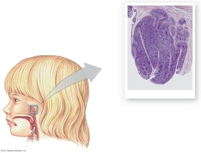
Tonsils: Lymphoid nodules in the pharynx; include palatine, pharyngeal, and lingual tonsils.
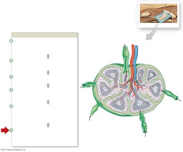
Lymph Nodes: Filter lymph and remove pathogens before returning fluid to the bloodstream.
The Thymus
The thymus is a bilobed gland located in the mediastinum, essential for T cell development. It is largest before puberty and shrinks with age, which may increase susceptibility to disease.
Structure: Divided into lobules containing a cortex (site of T cell maturation) and medulla (site of mature T cells).
Function: Produces thymosins, hormones necessary for T cell development.
The Spleen
The spleen is the largest lymphoid organ, located in the upper left abdomen. It filters blood, removes abnormal blood cells, stores iron, and initiates immune responses to blood-borne antigens.
Structure: Surrounded by a capsule, contains red pulp (RBCs) and white pulp (lymphocytes, macrophages).
Functions:
Removes abnormal blood components.
Stores iron.
Initiates immune responses by B and T cells.
Nonspecific (Innate) Defenses
Nonspecific defenses are present from birth and provide general protection against a wide range of pathogens. They include physical barriers, phagocytes, immunological surveillance, interferons, the complement system, inflammation, and fever.
Physical Barriers: Skin, mucous membranes, secretions, and hair.
Phagocytes: Neutrophils, eosinophils, and macrophages that engulf and destroy pathogens.
Immunological Surveillance: NK cells monitor and destroy abnormal cells.
Interferons: Proteins that interfere with viral replication and activate immune cells.
Complement System: Plasma proteins that enhance immune responses and destroy pathogens.
Inflammation: Localized response to injury or infection, characterized by swelling, heat, redness, and pain.
Fever: Elevated body temperature that inhibits pathogens and accelerates immune responses.
Specific (Adaptive) Defenses
Specific defenses involve the activities of T cells and B cells, which provide immunity against particular antigens. These responses are characterized by specificity, versatility, memory, and tolerance.
T cells: Provide cell-mediated immunity, targeting infected or abnormal cells.
B cells: Provide antibody-mediated immunity, targeting antigens in body fluids.
Properties of Immunity:
Specificity: Each lymphocyte recognizes one antigen.
Versatility: Millions of lymphocytes for different antigens.
Memory: Memory cells respond rapidly to previously encountered antigens.
Tolerance: Immune system ignores normal self-tissues.
Antigen Presentation and Activation of Lymphocytes
Antigen presentation is the process by which antigens are displayed on cell surfaces by MHC proteins, triggering specific immune responses.
MHC Class I: Present on all cells; display antigens from intracellular pathogens.
MHC Class II: Present on antigen-presenting cells; display antigens from extracellular pathogens.
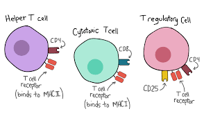
CD8 T cells: Recognize antigens on MHC I; differentiate into cytotoxic, memory, and suppressor T cells.
CD4 T cells: Recognize antigens on MHC II; help activate B cells and other immune cells.
Antibodies and Their Actions
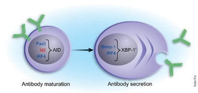
Antibodies are proteins produced by plasma cells (activated B cells) that bind to specific antigens and mediate their destruction through various mechanisms.
Structure: Composed of two heavy and two light chains, with variable regions forming antigen-binding sites.
Classes: IgG, IgE, IgD, IgM, IgA—each with distinct roles in immunity.
Mechanisms: Neutralization, prevention of pathogen adhesion, activation of complement, stimulation of inflammation, opsonization, attraction of phagocytes, precipitation, and agglutination.
Allergies and Immune Disorders
Allergies are inappropriate or excessive immune responses to antigens (allergens), often mediated by IgE antibodies. Reactions can be localized (e.g., hay fever) or systemic (e.g., anaphylaxis).
Integration of Nonspecific and Specific Defenses
Both branches of the immune system work together to protect the body. Nonspecific defenses provide immediate, broad protection, while specific defenses target particular threats and provide long-lasting immunity.
Summary Table: Major Lymphatic Organs and Functions
Organ | Location | Main Function |
|---|---|---|
Lymph nodes | Throughout body, concentrated in neck, axillae, groin | Filter lymph, house lymphocytes |
Tonsils | Pharynx | Trap pathogens entering through mouth/nose |
Thymus | Mediastinum | T cell maturation |
Spleen | Upper left abdomen | Filter blood, immune surveillance |
Bone marrow | Medullary cavity of bones | Production of lymphocytes |
