 Back
BackThe Lymphatic System and Immunity: Structure, Function, and Defense Mechanisms
Study Guide - Smart Notes
 Tailored notes based on your materials, expanded with key definitions, examples, and context.
The Lymphatic System and Immunity
Introduction
The lymphatic system is a crucial component of the body's defense mechanisms, working closely with the immune system to protect against disease and maintain fluid balance. This system consists of a network of vessels, tissues, and organs that transport lymph and house immune cells. Understanding its structure and function is essential for comprehending how the body defends itself from pathogens and maintains homeostasis.
Structure and Function of the Lymphatic System
Major Functions of the Lymphatic System
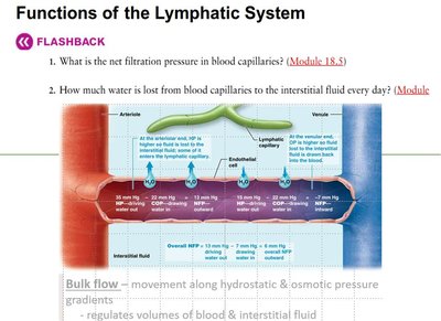
Fluid Recovery: Drains excess interstitial fluid and leaked plasma proteins from tissues back to the bloodstream, preventing edema.
Lipid Absorption: Transports dietary fats and fat-soluble vitamins from the digestive tract to the blood via specialized lymphatic vessels called lacteals.
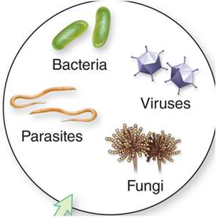
Immune Defense: Provides sites for immune surveillance and response, filtering lymph and blood for pathogens and foreign substances.
Comparison of Lymphatic Vessels and Blood Vessels
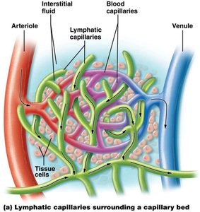
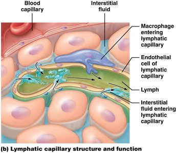
Lymphatic Capillaries: Begin as blind-ended tubes in tissues, more permeable than blood capillaries, allowing uptake of large molecules and particles.
Lymphatic Vessels: Have thinner walls and more valves than veins, ensuring one-way flow of lymph toward the heart.
Blood Vessels: Form a closed circuit, transporting blood under higher pressure; lymphatic vessels are part of an open system with lower pressure.
Pathway of Lymph Circulation
Lymphatic capillaries collect interstitial fluid, now called lymph.
Lymph flows through larger lymphatic vessels, passing through lymph nodes for filtration.
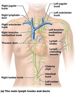
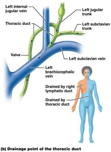
Principal lymphatic trunks (jugular, subclavian, bronchomediastinal, lumbar, intestinal) drain into two main ducts:
Right lymphatic duct: Drains right upper quadrant of the body into the right subclavian vein.
Thoracic duct: Drains the rest of the body into the left subclavian vein.
Mechanisms of Lymph Flow
Valves: Prevent backflow of lymph.
Skeletal Muscle Pump: Muscle contractions compress lymphatic vessels, propelling lymph forward.
Respiratory Pump: Pressure changes during breathing aid lymph movement toward the thoracic region.
Consequences of Lymphatic Dysfunction
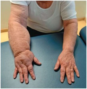
Edema: Accumulation of interstitial fluid due to impaired lymphatic drainage, leading to swelling.
Cellular Composition and Lymphoid Organs
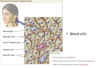
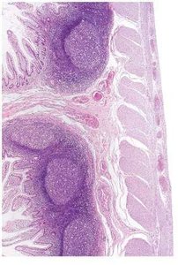
Microscopic Structure of Lymphoid Tissue
Reticular Connective Tissue: Forms the structural framework of lymphoid organs, supporting immune cells.
Cells Present: Macrophages, dendritic cells, B and T lymphocytes, reticular cells.
Primary Lymphoid Organs
Red Bone Marrow: Site of hematopoiesis; produces all blood cells, including lymphocytes.
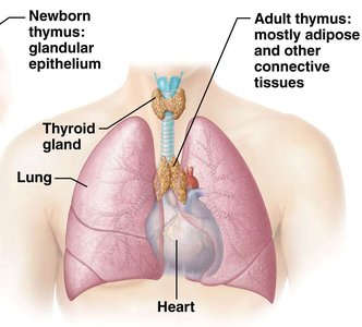
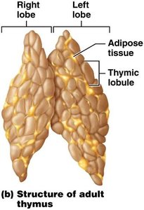
Thymus: Site of T lymphocyte maturation; large in children, atrophies with age.

Secondary Lymphoid Organs
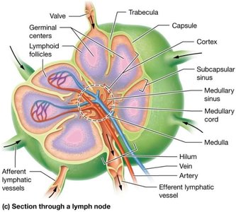
Lymph Nodes: Encapsulated structures located along lymphatic vessels; filter lymph and house immune cells.
Spleen: Largest lymphoid organ; filters blood, removes old erythrocytes, and mounts immune responses.
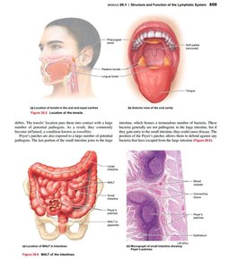
Mucosa-Associated Lymphoid Tissue (MALT): Includes tonsils, Peyer's patches, and appendix; protects mucosal surfaces.
Overview of the Immune System
Innate vs. Adaptive Immunity
Innate Immunity: Non-specific, present at birth, includes barriers and internal defenses (e.g., phagocytes, inflammation).
Adaptive Immunity: Specific, develops after exposure, involves lymphocytes (B and T cells) and memory formation.
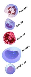
Cells of the Immune System
Phagocytes: Neutrophils, macrophages; ingest and destroy pathogens.
Lymphocytes: B cells (produce antibodies), T cells (cell-mediated immunity), NK cells (kill abnormal cells).
Other Cells: Dendritic cells (antigen presentation), mast cells (inflammation), basophils, eosinophils.
Innate Immunity: First and Second Lines of Defense
First Line of Defense: Surface Barriers
Physical Barriers: Skin and mucous membranes prevent pathogen entry.
Chemical Barriers: Secretions (tears, saliva, sweat, sebum, gastric juice) contain antimicrobial substances.
Mechanical Barriers: Actions like urination, defecation, and vomiting expel microbes.
Second Line of Defense: Internal Defenses
Antimicrobial Proteins: Interferons (inhibit viral replication), complement proteins (enhance immune response).
Natural Killer (NK) Cells: Destroy infected or abnormal cells by releasing perforins and granzymes.
Phagocytosis: Neutrophils and macrophages ingest and digest pathogens in three phases: chemotaxis, adherence, ingestion.
Inflammation: Local response to injury or infection; characterized by redness, heat, swelling, and pain. Stages include vasodilation, phagocyte migration, and tissue repair.
Fever: Elevated body temperature inhibits microbial growth and enhances tissue repair.
Adaptive Immunity: Third Line of Defense
Cell-Mediated Immunity
T Lymphocytes: Mature in the thymus; include helper T cells (Th, CD4+) and cytotoxic T cells (Tc, CD8+).
Activation: Requires antigen presentation by MHC molecules and costimulation.
Function: Tc cells destroy infected cells by inducing apoptosis or cytolysis; Th cells secrete cytokines to activate other immune cells.
Antibody-Mediated (Humoral) Immunity
B Lymphocytes: Mature in bone marrow; activated by antigen binding and helper T cell signals.
Plasma Cells: Differentiated B cells that secrete antibodies specific to the antigen.
Memory B Cells: Provide rapid response upon re-exposure to the same antigen.
Antibody Structure and Classes
Structure: Y-shaped molecule with two heavy and two light chains; variable region binds antigen, constant region determines class.
Classes:
IgG: Most abundant, crosses placenta.
IgA: Found in secretions, provides localized protection.
IgM: First antibody produced, involved in blood typing.
IgE: Involved in allergic responses.
IgD: Functions in B cell activation.
Immunological Memory
Primary Response: First exposure to antigen; slower and weaker antibody production.
Secondary Response: Subsequent exposures; faster and stronger due to memory cells, primarily IgG.
Summary Table: Key Lymphatic Organs and Functions
Organ | Location | Main Function |
|---|---|---|
Thymus | Thoracic cavity, above heart | T cell maturation |
Lymph Nodes | Along lymphatic vessels | Filter lymph, immune surveillance |
Spleen | Left upper quadrant, abdomen | Filters blood, recycles RBCs, immune response |
MALT | Mucosal linings (tonsils, Peyer's patches, appendix) | Protects mucosal surfaces from pathogens |
Additional info: The lymphatic and immune systems are closely integrated, with lymphoid organs providing sites for immune cell development, activation, and response. Disorders of the lymphatic system can lead to immune deficiencies, chronic infections, or autoimmune diseases.
