 Back
The Lymphatic System, Lymphoid Organs, and Immune System: Structure and Function
Study Guide - Smart Notes
 Tailored notes based on your materials, expanded with key definitions, examples, and context.
The Lymphatic System and Lymphoid Organs
Overview of the Lymphatic System
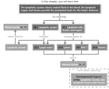
The lymphatic system returns leaked fluid from blood vessels back to the blood and provides the anatomical basis for the body's defenses. It consists of lymphatic vessels, lymph (the fluid), and lymphoid organs and tissues, which house immune cells.
Lymphatic vessels: Network that drains interstitial fluid and returns it to the bloodstream.
Lymph: Fluid collected from tissues, containing proteins, waste, and immune cells.
Lymph nodes: Filter lymph and house lymphocytes for immune surveillance.
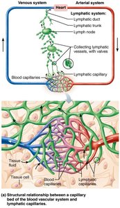
Structure and Distribution of Lymphatic Vessels
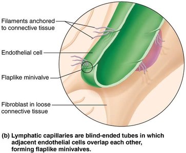
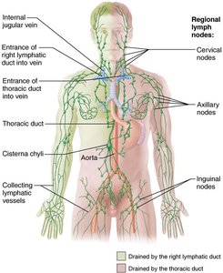
Lymphatic vessels form a one-way system that ensures lymph flows only toward the heart. The system begins with highly permeable lymphatic capillaries, which are found between tissue cells and blood capillaries in loose connective tissue. These capillaries are blind-ended tubes with overlapping endothelial cells that form flaplike minivalves, allowing large molecules (such as proteins and cell debris) to enter easily.
Lymphatic capillaries: Absent from bones, teeth, and bone marrow; present in meninges of the CNS.
Lacteals: Specialized lymphatic capillaries in the intestinal mucosa that absorb digested fats (chyle).
Collecting lymphatic vessels: Larger vessels with valves, similar to veins, that transport lymph to lymphatic trunks and ducts.
Major Lymphatic Trunks and Ducts
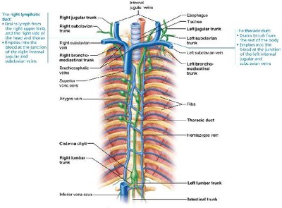
Lymphatic trunks drain large regions of the body and empty into two main ducts:
Right lymphatic duct: Drains right upper arm and right side of head and thorax.
Thoracic duct: Drains the rest of the body, beginning as the cisterna chyli.
Both ducts empty lymph into the venous circulation at the junction of the internal jugular and subclavian veins.
Lymphoid Cells and Tissues
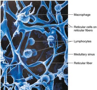
Lymphoid tissues are composed of immune system cells and supporting cells. The main immune cells are lymphocytes (T cells and B cells), which protect against antigens. Other important cells include macrophages (phagocytosis and activation of T cells), dendritic cells (antigen capture and presentation), and reticular cells (produce the stroma, a network of reticular fibers supporting immune cells).
Diffuse lymphoid tissue: Loose arrangement of lymphoid cells, found in most organs.
Lymphoid follicles (nodules): Solid, spherical bodies with germinal centers of proliferating B cells; found in lymph nodes, Peyer's patches, and appendix.
Lymphoid Organs
Lymph Nodes
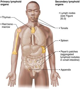
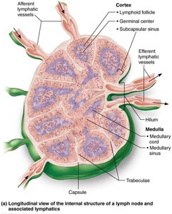
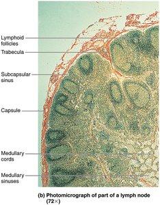
Lymph nodes are the principal secondary lymphoid organs, acting as filters for lymph and sites for immune activation. They are bean-shaped, less than 2.5 cm, and found in clusters along lymphatic vessels (notably in the inguinal, axillary, and cervical regions).
Structure: Surrounded by a capsule, divided into cortex (with follicles and germinal centers) and medulla (with medullary cords and sinuses).
Function: Cleansing lymph (macrophages remove debris) and immune activation (lymphocytes mount responses to antigens).
Spleen
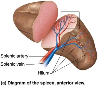
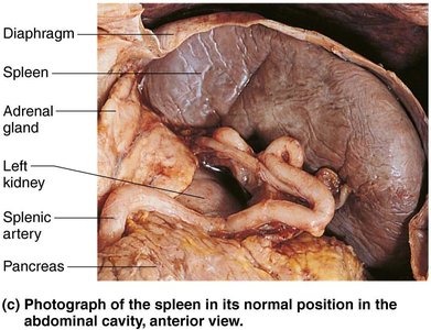
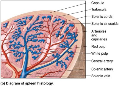
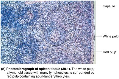
The spleen is the largest lymphoid organ, located in the left upper abdomen. It filters blood, removes old red blood cells and platelets, and provides a site for lymphocyte proliferation and immune surveillance.
White pulp: Site of immune function, rich in lymphocytes.
Red pulp: Site of destruction of old RBCs and pathogens, rich in macrophages.
Additional functions: Stores breakdown products of RBCs, platelets, and monocytes; may produce erythrocytes in the fetus.
MALT (Mucosa-Associated Lymphoid Tissue)
MALT consists of lymphoid tissues in mucous membranes throughout the body, including the tonsils, Peyer's patches (in the small intestine), and the appendix. These tissues protect against pathogens entering through mucosal surfaces.
Tonsils: Form a ring around the pharynx, trap and destroy pathogens in food and air.
Peyer's patches: Clusters of lymphoid follicles in the small intestine, prevent bacterial breach of the intestinal wall.
Appendix: Contains lymphoid follicles, aids in immune function in the gut.
Thymus
The thymus is a primary lymphoid organ where T lymphocytes mature. It is most active during childhood and gradually atrophies with age. The thymus differs from other lymphoid organs as it lacks B cells, does not directly fight antigens, and its stroma is made of epithelial cells.
Structure: Lobules with outer cortex (rapidly dividing lymphocytes) and inner medulla (fewer lymphocytes, thymic corpuscles).
Function: Secretes hormones for T cell maturation and immunocompetence.
The Immune System: Innate and Adaptive Defenses
Overview of Immune Defenses
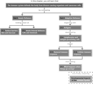
The immune system protects the body from disease-causing organisms and cancerous cells through two main defense systems: innate (nonspecific) and adaptive (specific) immunity.
Innate defenses: First and second lines of defense (surface barriers and internal cells/chemicals).
Adaptive defenses: Third line of defense, targeting specific antigens with memory.
Innate Defenses
Surface Barriers
Skin and mucous membranes provide a physical barrier to most microorganisms. Their secretions (acid, enzymes, mucin, defensins) inhibit or destroy pathogens.
Acid mantle: Inhibits bacterial growth.
Lysozyme: Enzyme that destroys bacteria.
Mucin: Traps microorganisms in mucus.
Defensins: Antimicrobial peptides.
Internal Defenses
If pathogens breach surface barriers, internal defenses are activated:
Phagocytes: Neutrophils and macrophages ingest and destroy pathogens.
Natural killer (NK) cells: Lyse cancer and virus-infected cells.
Inflammation: Local response to injury, characterized by redness, heat, swelling, and pain.
Antimicrobial proteins: Interferons (inhibit viral replication) and complement (enhances inflammation, promotes cell lysis).
Fever: Systemic response to infection, increases metabolic rate and inhibits microbial growth.
Adaptive Defenses
Adaptive immunity is specific, systemic, and has memory. It consists of humoral (antibody-mediated) and cellular (cell-mediated) responses.
Antigens: Substances that trigger adaptive immune responses; complete antigens have immunogenicity and reactivity.
Lymphocytes: B cells (produce antibodies) and T cells (directly attack infected cells).
Antigen-presenting cells (APCs): Dendritic cells, macrophages, and B cells that present antigens to T cells.
Humoral Immunity
B cells are activated by antigen binding, undergo clonal selection, and differentiate into plasma cells (secrete antibodies) and memory cells. Antibodies neutralize, agglutinate, precipitate antigens, and activate complement.
Cellular Immunity
T cells are activated by antigen presentation and co-stimulation. CD4 cells become helper T cells (activate B cells, T cells, and macrophages), while CD8 cells become cytotoxic T cells (destroy infected or abnormal cells). Regulatory T cells moderate immune responses.
Immune Disorders
Immunodeficiencies: Congenital or acquired conditions impairing immune function (e.g., SCID, AIDS).
Autoimmune diseases: Immune system attacks self-antigens (e.g., rheumatoid arthritis, type 1 diabetes).
Hypersensitivities: Overactive immune responses to harmless antigens (allergies, cytotoxic, immune complex, delayed types).
Developmental Aspects
Lymphatic vessels and lymphoid organs develop from embryonic veins and mesenchyme, except the thymus (endodermal origin). The immune system matures after birth, with efficiency declining with age.
