 Back
BackThe Muscular System: Structure, Function, and Organization
Study Guide - Smart Notes
 Tailored notes based on your materials, expanded with key definitions, examples, and context.
The Muscular System
Introduction to the Muscular System
The muscular system consists exclusively of skeletal muscles and is responsible for producing movement, maintaining posture, and supporting bodily functions. The organization of muscles, including the arrangement of muscle fibers (fascicles), significantly influences the power, range, and speed of movement.
Fascicle Arrangement in Skeletal Muscles
Types of Fascicle Arrangements
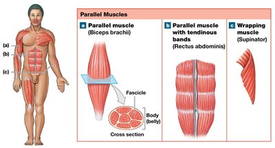
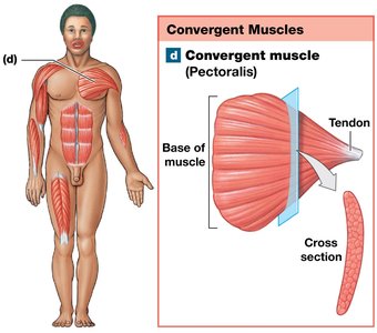
Skeletal muscle fibers are grouped into bundles called fascicles. The arrangement of these fascicles determines the muscle's shape and functional properties. The main types of fascicle arrangements are:
Parallel muscles: Fascicles are parallel to the long axis of the muscle. These muscles can be flat or spindle-shaped (fusiform) with a central body (belly). They can shorten significantly during contraction. Examples: rectus abdominis, biceps brachii.
Convergent muscles: Fascicles spread over a broad area and converge at a common attachment site (tendon, aponeurosis, or raphe). These muscles can pull in different directions. Example: pectoralis major.
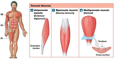
Pennate muscles: Fascicles form an angle with the tendon. These muscles contain more fibers and generate more tension but have a shorter range of motion. Types include:
Unipennate: All fascicles on the same side of the tendon (extensor digitorum).
Bipennate: Fascicles on both sides of a central tendon (rectus femoris).
Multipennate: Tendon branches within the muscle (deltoid).
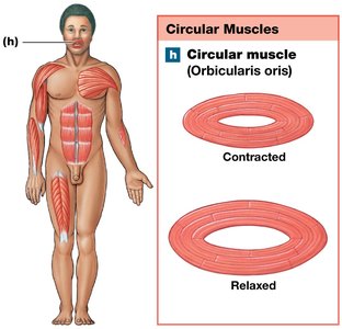
Circular muscles (sphincters): Fascicles are concentrically arranged around an opening, acting as valves to open and close passages. Example: orbicularis oris (mouth).
Levers and Muscle Efficiency
Classes of Levers in the Body
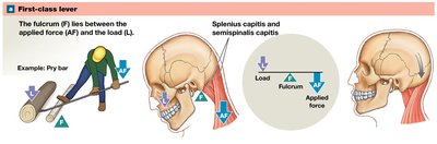
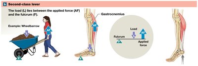
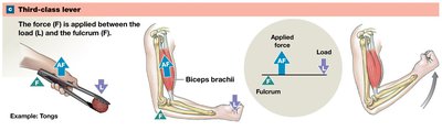
Muscles attach to bones and create movement by acting as levers. A lever is a rigid structure that moves around a fixed point called the fulcrum. The force applied by muscles is called the applied force (AF), which acts against a load (L). Levers can change the direction, distance, speed, and strength of movement. There are three classes of levers:
First-class lever: The fulcrum is between the applied force and the load (like a seesaw). Example: Extension of the neck and lifting the head.
Second-class lever: The load is between the applied force and the fulcrum (like a wheelbarrow). Example: Plantar flexion at the ankle by the calf muscles.
Third-class lever: The applied force is between the load and the fulcrum (like tweezers). This is the most common lever in the body and maximizes speed and distance. Example: Elbow flexion by the biceps brachii.
Muscle Origins, Insertions, and Actions
Definitions and Functional Groups
Muscles produce movement by contracting and pulling on bones at specific attachment points:
Origin: The fixed (less movable) attachment point, usually proximal.
Insertion: The more movable attachment point, usually distal.
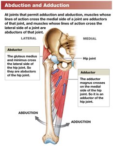
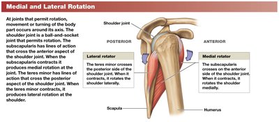
Action: The specific movement produced by muscle contraction (e.g., flexion, extension, abduction).
Muscles work together in groups to maximize efficiency:
Agonist (prime mover): Main muscle responsible for a movement.
Antagonist: Opposes the action of the agonist.
Synergist: Assists the agonist.
Fixator: Stabilizes the origin of the agonist.
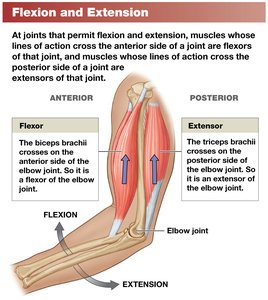
Agonists and antagonists work in pairs, such as flexors and extensors or abductors and adductors.
Naming Skeletal Muscles
Muscle Naming Conventions
The names of skeletal muscles provide information about their location, structure, and function. Common naming criteria include:
Region of the body: e.g., temporalis (temple), brachialis (arm).
Position, direction, or fascicle arrangement: e.g., externus (superficial), rectus (straight), oblique (slanting).
Structural characteristics: e.g., number of tendons (biceps = two heads), shape (deltoid = triangle), size (maximus = largest).
Action: e.g., flexor (bending), extensor (straightening), abductor (movement away from midline).
Understanding these conventions helps in identifying the muscle's location, appearance, or function.
Axial and Appendicular Muscles
Divisions of the Muscular System
The muscular system is divided into two main groups:
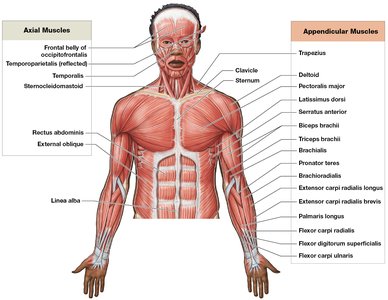
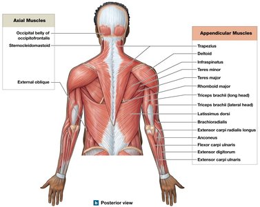
Axial muscles: Position the axial skeleton, including the head, vertebral column, and rib cage. They make up about 60% of skeletal muscles.
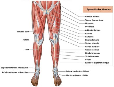
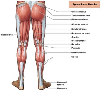
Appendicular muscles: Support and move the appendicular skeleton (limbs and girdles).
Principal Axial Muscles
Muscles of the Head and Neck
Axial muscles are grouped by location and function:
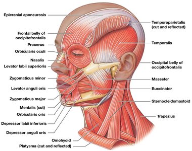
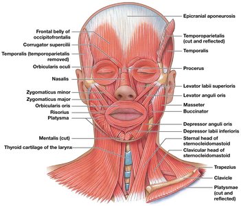
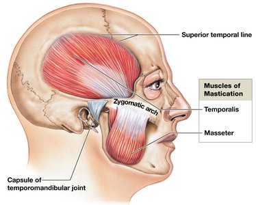
Muscles of facial expression: orbicularis oris (purses lips), buccinator (compresses cheeks), zygomaticus major (smile), orbicularis oculi (closes eyes).
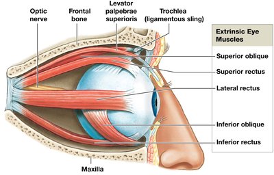
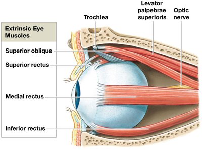
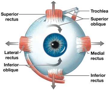
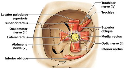
Extrinsic eye muscles: Move the eyeball in various directions (e.g., superior rectus, lateral rectus).
Muscles of mastication: masseter and temporalis (elevate mandible), pterygoid muscles (side-to-side movement).
Muscles of the tongue: genioglossus, hyoglossus, styloglossus, palatoglossus.
Muscles of the pharynx: pharyngeal constrictors, palatal muscles, laryngeal elevators.
Muscles of the anterior neck: digastric, mylohyoid, sternocleidomastoid.
Muscles of the Vertebral Column
These muscles stabilize, flex, extend, and rotate the vertebral column. Major groups include:
Erector spinae: spinalis, longissimus, iliocostalis (extension and lateral flexion).
Spinal flexors: longus capitis, longus colli, quadratus lumborum.
Oblique and Rectus Muscles
These muscles compress the abdomen, flex and rotate the vertebral column, and assist in breathing:
Oblique muscles: external oblique, internal oblique, transversus abdominis.
Rectus muscles: rectus abdominis (flexes vertebral column, compresses abdomen).
Diaphragm: Main muscle of respiration, separates thoracic and abdominopelvic cavities.
Muscles of the Pelvic Floor
These muscles support pelvic organs, control openings of the urethra and anus, and stabilize the pelvic floor:
Pelvic diaphragm: levator ani, coccygeus.
External urethral sphincter and external anal sphincter: Control passage of urine and feces.
Principal Appendicular Muscles
Muscles of the Shoulders and Upper Limbs
Muscles that position the pectoral girdle: trapezius, serratus anterior, pectoralis minor, rhomboids, levator scapulae.
Muscles that move the arm: deltoid, pectoralis major, latissimus dorsi, rotator cuff muscles (supraspinatus, infraspinatus, teres minor, subscapularis).
Muscles that move the forearm and hand: biceps brachii, triceps brachii, brachialis, brachioradialis, pronator teres, supinator.
Muscles that move the wrist, hand, and fingers: flexor carpi radialis, flexor carpi ulnaris, extensor carpi radialis, extensor carpi ulnaris, extensor digitorum, flexor digitorum.
Intrinsic muscles of the hand: Responsible for fine motor movements (e.g., adductor pollicis, opponens pollicis).
Muscles of the Pelvis and Lower Limbs
Muscles that move the thigh: gluteus maximus, gluteus medius, tensor fasciae latae, adductor group, iliopsoas group.
Muscles that move the leg: hamstrings (biceps femoris, semitendinosus, semimembranosus), quadriceps femoris (rectus femoris, vastus lateralis, vastus medialis, vastus intermedius).
Muscles that move the foot and toes: gastrocnemius, soleus, tibialis anterior, extensor digitorum longus, flexor digitorum longus.
Intrinsic muscles of the foot: Maintain arches and move the toes.
Functional Relationships and Effects of Exercise
Integration with Other Body Systems
The muscular system works closely with other body systems:
Cardiovascular system: Delivers oxygen and nutrients, removes waste.
Respiratory system: Meets increased oxygen demand during muscle activity.
Integumentary system: Disperses heat generated by muscles.
Nervous and endocrine systems: Regulate and coordinate muscle activity.
Exercise enhances muscle performance and has systemic effects, improving cardiovascular health, respiratory efficiency, and metabolic function.
Additional info: For a comprehensive understanding, students should also review the innervation of major muscles, the role of muscle tone in posture, and the adaptations of muscle tissue to different types of exercise (e.g., endurance vs. resistance training).
