 Back
BackThe Nervous System: Structure and Function
Study Guide - Smart Notes
 Tailored notes based on your materials, expanded with key definitions, examples, and context.
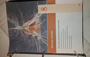
The Nervous System
Introduction to the Nervous System
The nervous system is the master controlling and communicating system of the body. Every thought, action, and emotion reflects its activity. The nervous system uses electrical impulses to interpret and respond to what is happening both inside and outside the body.
Sensory Input: Gathering information from sensory receptors about internal and external changes.
Integration: Processing and interpreting sensory input and deciding what should be done at each moment.
Motor Output: The response by activating effector organs (muscles or glands).
Organization of the Nervous System
Structural and Functional Classification
The nervous system is divided into two main parts: the central nervous system (CNS) and the peripheral nervous system (PNS). Each part has distinct structures and functions.
Central Nervous System (CNS): Consists of the brain and spinal cord. It is the integrating and command center.
Peripheral Nervous System (PNS): Consists of nerves that extend from the brain and spinal cord. It serves as communication lines linking all parts of the body to the CNS.
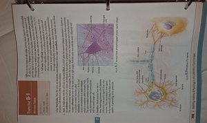
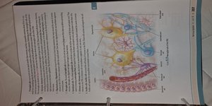
Cells of the Nervous System
The nervous system is composed of two main types of cells: neurons and neuroglia (supporting cells).
Neurons: The functional units of the nervous system, specialized to transmit messages.
Neuroglia: Supporting cells that protect, insulate, and support neurons.
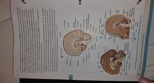
Anatomy of the Brain
Major Regions of the Brain
The brain is divided into several major regions, each with specialized functions. These include the cerebrum, diencephalon, brain stem, and cerebellum.
Cerebrum: The largest part, responsible for voluntary activities, intelligence, memory, and sensory processing.
Diencephalon: Contains structures such as the thalamus and hypothalamus, involved in sensory relay and homeostasis.
Brain Stem: Controls basic life functions such as breathing and heart rate.
Cerebellum: Coordinates muscle movements and maintains posture and balance.
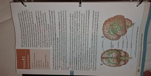
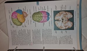
Lobes of the Cerebrum
The cerebrum is divided into lobes, each associated with different functions.
Frontal Lobe: Controls voluntary movement, reasoning, planning, and problem-solving.
Parietal Lobe: Processes sensory information such as touch, temperature, and pain.
Temporal Lobe: Involved in hearing, memory, and speech.
Occipital Lobe: Responsible for visual processing.
Internal Structures of the Brain
Internal brain structures include the thalamus, hypothalamus, pituitary gland, and brain stem components. These structures are essential for sensory processing, hormone regulation, and autonomic functions.
Thalamus: Relay station for sensory impulses.
Hypothalamus: Regulates body temperature, hunger, thirst, and other homeostatic systems.
Brain Stem: Includes the midbrain, pons, and medulla oblongata.
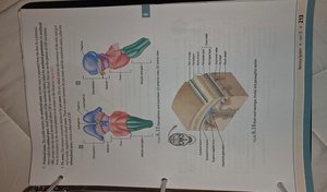
Protection of the Central Nervous System
Meninges and Cerebrospinal Fluid
The brain and spinal cord are protected by three connective tissue membranes called meninges, as well as by cerebrospinal fluid (CSF) which cushions the CNS.
Dura Mater: The tough outermost layer.
Arachnoid Mater: The middle, web-like layer.
Pia Mater: The delicate innermost layer, closely attached to the brain and spinal cord.
Cerebrospinal Fluid (CSF): Circulates in the subarachnoid space, providing a protective cushion.
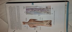
The Spinal Cord
Structure and Function
The spinal cord is a cylindrical structure that extends from the brain stem to the lower back. It serves as a major pathway for information traveling between the brain and the rest of the body.
Gray Matter: Contains neuron cell bodies and is involved in processing information.
White Matter: Contains myelinated nerve fibers that transmit signals up and down the spinal cord.
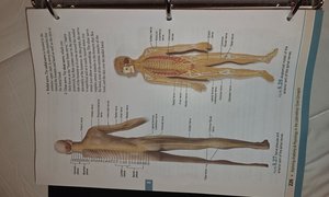
Spinal Nerves: 31 pairs of nerves that emerge from the spinal cord to innervate the body.
Peripheral Nervous System (PNS)
Nerves and Ganglia
The PNS consists of cranial and spinal nerves, as well as ganglia (clusters of neuron cell bodies outside the CNS). It connects the CNS to limbs and organs.
Cranial Nerves: 12 pairs that emerge from the brain and primarily serve the head and neck.
Spinal Nerves: 31 pairs that emerge from the spinal cord and serve the rest of the body.
Ganglia: Collections of neuron cell bodies located outside the CNS.
Functional Divisions of the PNS
The PNS is functionally divided into the sensory (afferent) division and the motor (efferent) division. The motor division is further subdivided into the somatic and autonomic nervous systems.
Sensory (Afferent) Division: Transmits impulses from sensory receptors to the CNS.
Motor (Efferent) Division: Transmits impulses from the CNS to effector organs.
Somatic Nervous System: Controls voluntary movements of skeletal muscles.
Autonomic Nervous System: Regulates involuntary activities of smooth muscle, cardiac muscle, and glands.
Summary Table: Major Divisions of the Nervous System
Division | Main Structures | Primary Function |
|---|---|---|
Central Nervous System (CNS) | Brain, Spinal Cord | Integration, command center |
Peripheral Nervous System (PNS) | Cranial nerves, Spinal nerves, Ganglia | Communication lines between CNS and body |
Somatic Nervous System | Motor neurons to skeletal muscle | Voluntary control of body movements |
Autonomic Nervous System | Motor neurons to smooth/cardiac muscle, glands | Involuntary control of visceral functions |
Key Terms and Definitions
Neuron: A nerve cell; the basic building block of the nervous system.
Synapse: The junction between two neurons where information is transmitted.
Action Potential: An electrical impulse that travels along the membrane of a neuron.
Reflex: A rapid, automatic response to a stimulus.
Example: Reflex Arc
A reflex arc is the simplest type of nerve pathway, involving a sensory neuron, an interneuron, and a motor neuron. It allows for quick responses to stimuli without conscious thought.
Example: The knee-jerk reflex is a classic example of a simple reflex arc.
Additional Info
The nervous system is essential for maintaining homeostasis by detecting changes and initiating appropriate responses.
Damage to the nervous system can result in loss of sensation, movement, or cognitive function, depending on the area affected.
