 Back
BackThe Nervous System: Structure, Function, and Neurophysiology
Study Guide - Smart Notes
 Tailored notes based on your materials, expanded with key definitions, examples, and context.
11.1 The Nervous System
Overview and Functions
The nervous system is the master control and communication system of the body, capable of overriding all other body systems. It utilizes both electrical signals (within neurons) and chemical signals (neurotransmitters between neurons) for communication. Neurotransmitters are released from a presynaptic neuron, bind to a postsynaptic neuron, and can produce either excitatory or inhibitory effects.
Sensory Input: Collects information from internal and external environments as different stimuli.
Integration: Processes and interprets sensory input in the brain and spinal cord.
Motor Output: Activates effector organs (muscles and glands) to elicit a response.
Organization of the Nervous System
Central Nervous System (CNS): Consists of the brain and spinal cord, located in the dorsal body cavity.
Peripheral Nervous System (PNS): Composed of nerves (bundles of axons) outside the CNS, including 31 pairs of spinal nerves and 12 pairs of cranial nerves.
Divisions of the PNS
Sensory (Afferent) Division: Transmits impulses to the CNS from sensory receptors.
Somatic afferent fibers: From skin, skeletal muscles, and joints (frequent input).
Visceral afferent fibers: From organs (monitor baseline conditions).
Motor (Efferent) Division: Transmits impulses from the CNS to effector organs.
Somatic Nervous System: Voluntary control of skeletal muscles.
Autonomic Nervous System (ANS): Involuntary control of cardiac muscle, smooth muscle, and glands.
Sympathetic Division: "Fight or flight" responses.
Parasympathetic Division: "Rest and digest" responses.
11.2 Neuroglia
Types and Functions of Neuroglia
Neuroglia (glial cells) are support cells in the nervous system that provide structural and metabolic support to neurons, maintain the extracellular environment, and participate in defense mechanisms. The nervous system contains very little extracellular space due to the presence of these cells.
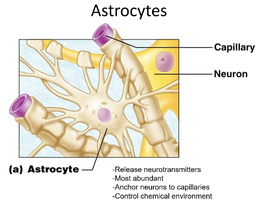
Astrocytes (CNS): Anchor neurons to blood supply, release neurotransmitters, help form the blood-brain barrier, and maintain the chemical environment.
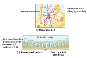
Microglia (CNS): Act as immune defense (phagocytes), removing pathogens and debris.
Ependymal Cells (CNS): Line brain and spinal cord cavities, circulate cerebrospinal fluid (CSF), and form a permeable barrier between CSF and tissue fluid.
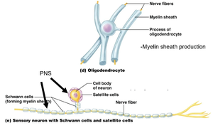
Oligodendrocytes (CNS): Form myelin sheaths around axons in the CNS.
Satellite Cells (PNS): Protect neuron cell bodies and regulate the environment around them.
Schwann Cells (PNS): Form myelin sheaths around axons in the PNS and protect neuron cell bodies.
11.3 Neurons
Structure and Function
Neurons are the functional units of the nervous system, specialized for the transmission of electrical impulses. They are primarily amitotic (do not divide), have a long lifespan, and require a constant supply of oxygen and glucose due to high energy demands.
Cell Body (Soma): Contains the nucleus and organelles, produces proteins and neurotransmitters, and lacks centrioles (cannot divide).
Lipofuscin: Pigment that accumulates with age, indicating aging neurons.
Myelination
Myelin Sheaths: Insulating layers around axons, composed of lipids and proteins, produced by oligodendrocytes (CNS) and Schwann cells (PNS). They speed up signal transmission.
Saltatory Conduction: In myelinated axons, the signal "jumps" between nodes of Ranvier, increasing conduction speed (~150 m/s).
Continuous Conduction: In unmyelinated axons, the signal travels slowly (0.5–10 m/s).
Nodes of Ranvier: Gaps between myelin sheaths where ion channels are concentrated and depolarization occurs.
Neuron Processes
Dendrites: Short, branched extensions that receive signals and convey them toward the cell body (graded potentials).
Axons: Long processes that transmit action potentials away from the cell body to axon terminals, where neurotransmitters are released.
Axon Terminals: Secretory regions that release neurotransmitters into the extracellular space.
Cytoskeletal System: Supports anterograde (away from cell body) and retrograde (toward cell body) transport of materials.
Structural Classification of Neurons
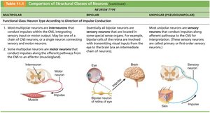
Multipolar: Three or more processes (one axon, multiple dendrites); most common in CNS.
Bipolar: Two processes (one axon, one dendrite); rare, found in special sensory organs.
Unipolar: Single short process that divides into peripheral and central processes; primarily sensory neurons in PNS.
Functional Classification of Neurons
Sensory (Afferent): Transmit impulses toward the CNS; primarily unipolar.
Motor (Efferent): Transmit impulses away from the CNS to effector organs; primarily multipolar.
Interneurons (Association Neurons): Integrate and connect sensory and motor pathways; 99% of all neurons; multipolar; located in CNS.
Neurophysiology
Neurons are excitable and generate action potentials in response to stimuli.
Ohm’s Law:
I: Current (flow of ions)
V: Voltage (potential difference)
R: Resistance (mainly due to plasma membrane)
11.4 The Resting Membrane Potential
Establishment and Maintenance
The resting membrane potential is the voltage difference across the neuron's membrane, typically ranging from -40 to -90 mV (inside negative). It is established by differences in ion concentrations and membrane permeability, maintained by the sodium/potassium pump (3 Na+ out, 2 K+ in). Potassium is the most important cation in this process.
Hyperpolarization: Occurs when potassium channels remain open, making the inside more negative and reducing the likelihood of a nerve impulse.
11.5 Graded Potentials
Characteristics and Types
Graded potentials are short-lived, local changes in membrane potential that can be excitatory or inhibitory. Their magnitude varies with stimulus strength and they decrease in intensity with distance due to the leaky plasma membrane. They occur in dendrites and soma, triggered by environmental stimuli, and can initiate action potentials if strong enough.
Receptor/Generator Potentials: Due to sensory receptor excitation.
Postsynaptic Potentials: Due to neurotransmitter binding.
11.6 Action Potentials
Generation and Propagation
Action potentials are brief reversals of membrane potential (total amplitude ~100 mV) generated by neurons and muscle cells. They do not decrease in strength over distance and are the principal means of neural communication. Once generated, action potentials follow the all-or-none principle and their frequency encodes stimulus intensity.
Absolute Refractory Period: Neuron cannot respond to another stimulus (Na+ channels open).
Relative Refractory Period: Follows absolute; strong stimulus can initiate another action potential (K+ channels open).
Conduction Velocity: Depends on axon diameter (larger = faster) and myelination (myelinated = faster, saltatory conduction).
Classification of Axons
Group | Diameter | Myelination | Speed | Function |
|---|---|---|---|---|
A | Large | Thick | 150 m/s | Somatic sensory/motor (skin, muscle, joints) |
B | Intermediate | Light | 15 m/s | Autonomic motor (viscera) |
C | Small | None | 1 m/s | Autonomic/slow pain, touch |
11.7 Synapses
Structure and Function
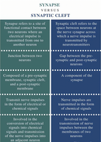
Synapses are specialized junctions that mediate information transfer between neurons. The synaptic cleft is the tiny gap where neurotransmitters diffuse. Synapses can be electrical (direct ion flow via connexons) or chemical (neurotransmitter-mediated).
Presynaptic Neuron: Conducts impulses toward the synapse.
Postsynaptic Neuron: Transmits impulses away from the synapse.
Axodendritic Synapses: Axon to dendrite.
Axosomatic Synapses: Axon to cell body.
Signal Flow Through a Neuron
Neurotransmitter released from presynaptic neuron
Crosses synaptic cleft
Binds to receptors on dendrites
Opens ion channels
Causes graded potential
If threshold reached at axon hillock, action potential generated
Travels down axon, neurotransmitter released again
Synaptic Delay and Termination
Synaptic Delay: Time required for neurotransmitter release, diffusion, and binding (0.3–0.5 ms); rate-limiting step.
Termination: Neurotransmitter is removed by enzymatic degradation, reuptake, or diffusion away from the synaptic cleft.
11.8 Postsynaptic Potentials
Types and Summation
Postsynaptic potentials are changes in membrane potential mediated by neurotransmitter receptors. The effect depends on the amount and duration of neurotransmitter binding.
Excitatory Postsynaptic Potentials (EPSPs): Graded potentials that depolarize the membrane, possibly initiating an action potential. Summation can be temporal (rapid-fire from one neuron) or spatial (multiple neurons firing simultaneously).
Inhibitory Postsynaptic Potentials (IPSPs): Hyperpolarize the membrane, making action potential generation less likely (increased K+ or Cl- permeability).
Synaptic Potentiation: Repeated use of a synapse increases its efficiency, enhancing communication (basis for learning and memory).
11.9 Neurotransmitters
Types and Functions
Neurotransmitters are chemicals used for communication within the nervous system. Over 50 have been identified and are classified by chemical structure and function.
Chemical Classes: Acetylcholine, biogenic amines, amino acids, peptides, ATP, and dissolved gases (NO, CO).
Functional Classification:
Effects: Excitatory or inhibitory (depends on receptor type).
Actions: Direct (binds and opens ion channels, fast) or indirect (acts via second messengers, slow and long-lasting).
Neuromodulators: Chemicals that modulate synaptic transmission without directly causing EPSPs or IPSPs; can influence neurotransmitter synthesis, release, degradation, or reuptake.
11.10 Neural Integration
Processing and Circuitry
Neural integration refers to the coordinated activity of groups of neurons to produce unified function. This is essential for complex behaviors and higher-level processing.
Patterns of Neural Processing
Serial Processing: Input travels along one pathway to a specific destination (e.g., spinal reflexes).
Parallel Processing: Input travels along multiple pathways, allowing for complex and simultaneous processing (important for higher mental functions).
Neural Circuits
Diverging: One input, many outputs (amplifies signal).
Converging: Many inputs, one output (concentrates signal).
Reverberating: Signal travels through a chain of neurons, each feeding back to previous neurons (oscillations, rhythmic activity).
Parallel After-Discharge: Input diverges to several chains, then converges to a single output (complex processing).
Neural Plasticity
Ability of neural circuits to change strength and connectivity (basis for learning and memory).
Peak during childhood, diminishes with age; exercise enhances neuroplasticity by increasing brain blood flow.
